 |
|
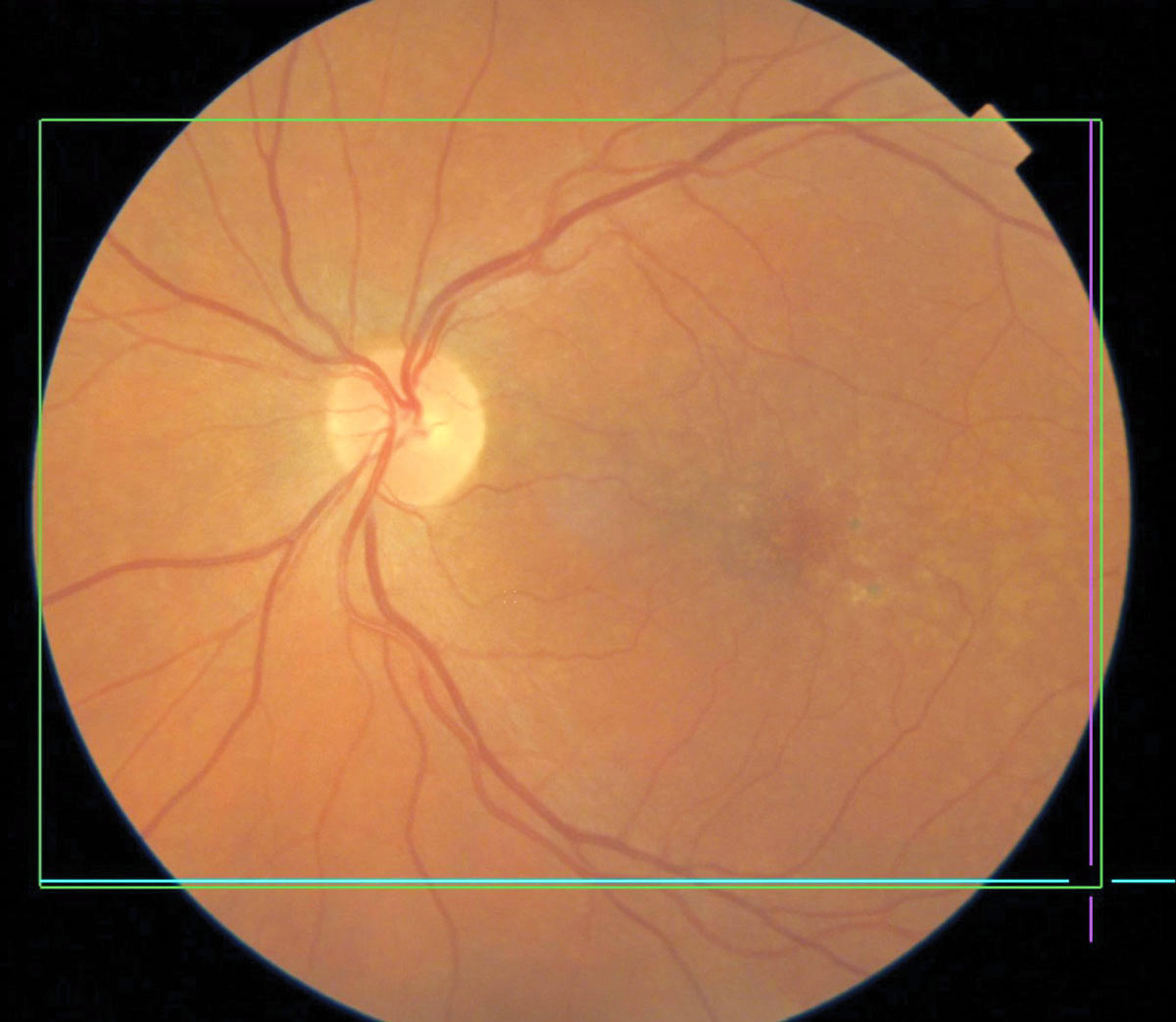
MNV lesion growth is detectable on OCT-A, which may help guide treatment for these patients. Photo: Brian Chou, OD. Click image to enlarge. |
The presence of treatment-naïve nonexudative macular neovascularization (MNV), which is detected by swept-source OCT angiography (OCT-A) before the onset of exudation in eyes with age-related macular degeneration (AMD), may predispose patients to the development of exudative changes. However, the duration from the nonexudative state to the onset of exudation in eyes with MNV varies. In a recent study, researchers aimed to investigate the growth of MNV in AMD.
A total of 45 patients with treatment-naïve nonexudative AMD in one eye and exudative AMD in the fellow eye who underwent OCT-A imaging for at least 12 months were retrospectively reviewed. The MNV area measurement was quantified in eyes with treatment-naïve nonexudative MNV using ImageJ to analyze the correlation between MNV growth and exudation onset and to evaluate the consistency of the MNV growth rate during the subclinical and exudative stages.
Treatment-naïve nonexudative MNV was identified in 21 of 45 fellow eyes from patients previously diagnosed with exudative AMD. Eyes with treatment-naïve nonexudative MNV showed a higher risk of developing exudation than eyes without. “In fact, MNV growth was commonly detected in treatment-naïve nonexudative MNV lesions; however, the lesion growth rate was diverse, ranging from -0.05% to 212.73%,” the study authors explained in their paper for the journal Eye. “Furthermore, the risk of exudation was greater for eyes with growing MNV than eyes with stable MNV.”
Among cases of subclinical nonexudative MNV in which exudation occurred, the sustained growth of MNV lesions was detected during anti-VEGF treatment. “These results suggest that the growth rate of MNV in the subclinical stage may indicate the innate activity of the neovascular lesions, which may be associated with exudation risks and may not be altered by initial anti-VEGF therapy presumably,” the team noted.
Based on the lesion area change in subclinical stage, the investigators subdivided the study eyes with subclinical MNV into stable MNV (less than 50% area growth or shrinkage) or unstable MNV (more than 50% area growth) to describe the biological activity of neovascular lesions.
“Eyes with unstable MNV correlated with more risks of exudation and developed exudation earlier than eyes with stable MNV,” they concluded. “Sustained growth of MNV was identified in unstable MNV lesions after they developed exudation despite ongoing anti-VEGF therapy. As an indicator of the MNV lesion activity, lesion growth might contribute to establishing treatment strategies and follow-up planning for AMD patients.”
Wang Y, Sun J, Jia H, et al. Growth of nonexudative macular neovascularization in age-related macular degeneration: an indicator of biological lesion activity. Eye. November 25, 2022. [Epub ahead of print]. |
