 |
|
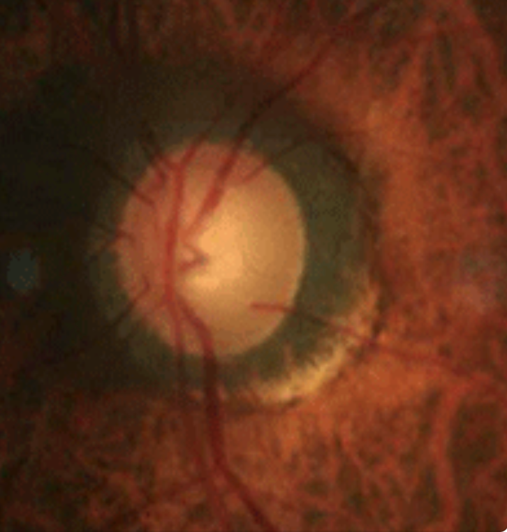
Parapapillary atrophy (seen here in a glaucoma patient), along with optic disc area increase, was more associated with age in the adolescent age group and with SER and AL in the young adult age group. Photo: Joseph W. Sowka, OD. Click image to enlarge. |
Many studies have shown that as myopia increases, so too do axial length (AL), parapapillary atrophy area and degree of optic disc, while the thickness and blood flow of the retina and choroid decrease. However, researchers in China believe that the optic discs of young high myopia patients remain largely unexplored in younger Asian populations. Their recent study quantitatively assessed the morphological and structural characteristics of the optic disc in young people with simple high myopia using OCT-A. They found that the parapapillary atrophy area, degree of optic disc tilt and disc area increased with AL and peripapillary retinal nerve fiber layer (RNFL) thickness but decreased with spherical equivalent refraction (SER). The association between these factors was slightly different in the adolescent (ages 12 to 17) and young adult (ages 18 to 30) groups. The degree of the optic disc tilt was more associated with AL and SER in the adolescent group, while disc area showed more of a correlation with AL and SER in the young adult group.
The cross-sectional study included a total of 112 patients with high myopia (SER ≤-6.00D) aged 12 to 30 years old. Parapapillary atrophy and ovality index from scanning laser ophthalmoscopy images and the degree of optic disc tilt from the optic nerve head were evaluated. OCT B-scans were analyzed as well. Peripapillary RNFL thickness and radial peripapillary capillary vessel density around the optic disc were obtained from the images of the optic disc angiography scan. The mean SER and axial length were -8.26D and 26.86mm, respectively. The mean parapapillary atrophy area, degree of the optic disc tilt and ovality index were 2.76mm2, 1.24 and 34.14, respectively.
The researchers proposed that parapapillary atrophy area might be caused by the stretching of the sclera and optic nerve during the progression of myopia. Therefore, the mechanisms underlying the change in parapapillary atrophy area among various age groups of high myopia need to be further validated.
The degree of optic disc tilt was associated with a more myopic SER in the adolescent group and with a longer AL in the young adult group. The researchers speculated that optic disc tilt may occur under the mechanism of scleral stretching by axial elongation during myopia progression among highly myopic people.
“It is novel that we observed the optic disc tilt was more associated with the AL and SER in the adolescent group, which might be the reason that the change in AL and SER was stronger in this age group,” they wrote in their paper.
The study concluded that monitoring the characteristics of the optic disc should be considered when managing patients with high myopia.
Zhang F, Liu X, Wang Y, et al. Characteristics of the optic disc in young people with high myopia. BMC Ophthalmol. 2022;22(1):477. |
