| The Optometric Retina Society can be reached via e-mail at optometric.retina.society@gmail.com or online at optometricretinasociety.org. |
FROM THE DESK OF THE EDITOR
Greetings. I am excited to start off our first newsletter of 2024 with a new look! The same retina-themed content remains, but a long overdue aesthetics update has arrived. The format of our newsletter has had little change since it was developed about 20 years ago. Twenty years is a long time. Just think how much eyecare has changed in the past 20 years. The therapeutics landscape has evolved from only prescribing topicals to allowing orals, injectables, and even some laser treatments. I’ve only been practicing optometry myself for 14 years, however, as a second-generation OD, I’ve been around optometry my whole life. I remember (early 2000s) hearing my father talk about taking continuing education classes in order to be able to begin prescribing orals. He considered that a big victory for the profession, and it was. For perspective, when he came into practice in the late 80s, he wasn’t legally allowed to dilate eyes, as his state license restricted use of topical diagnostics. Imaging, as well, has undergone a dramatic shift from fluorescein angiography, invented at Indiana University (my alma mater) in the 1960s, to now dyeless angiography via OCTA. In 20 years, we’ve gone through three different prototypes of OCT. The first time domain OCT rolled out for commercial sale in 1996. Since then, it has been replaced with spectral domain OCT and more recently swept source OCT. My point here, 20 years has seen a lot of change and thus a face-lift was long overdue. I hope you enjoy the new look as well as the content below. Cheers!
Anna Bedwell, OD, FAAO, FORS
Editor-in-Chief
Interested in a fellowship in the Optometric Retina Society? Click here for details regarding the application process.
YOU MAKE THE DIAGNOSIS
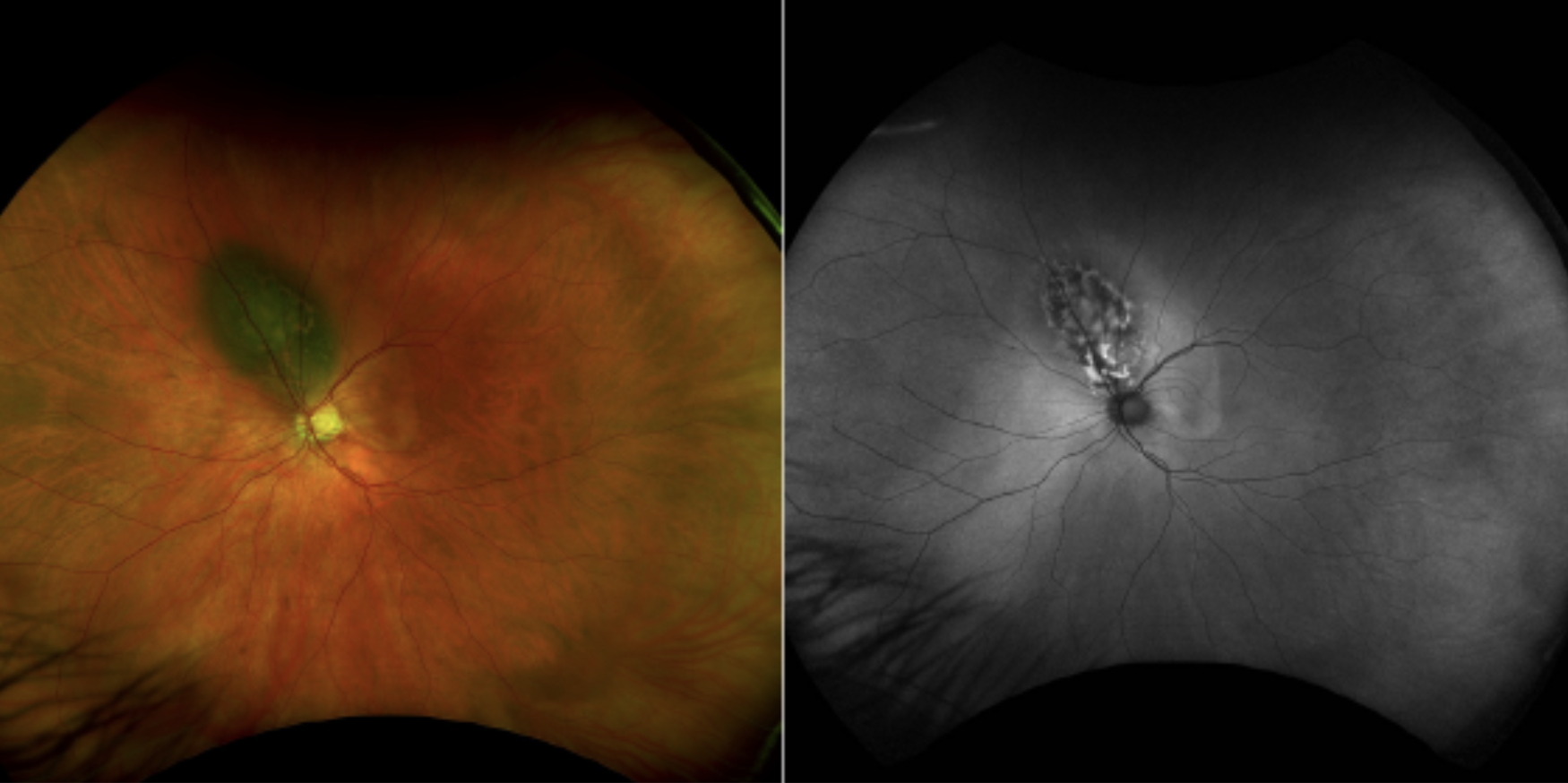 |
| Click image to enlarge. |
IMAGE GALLERY
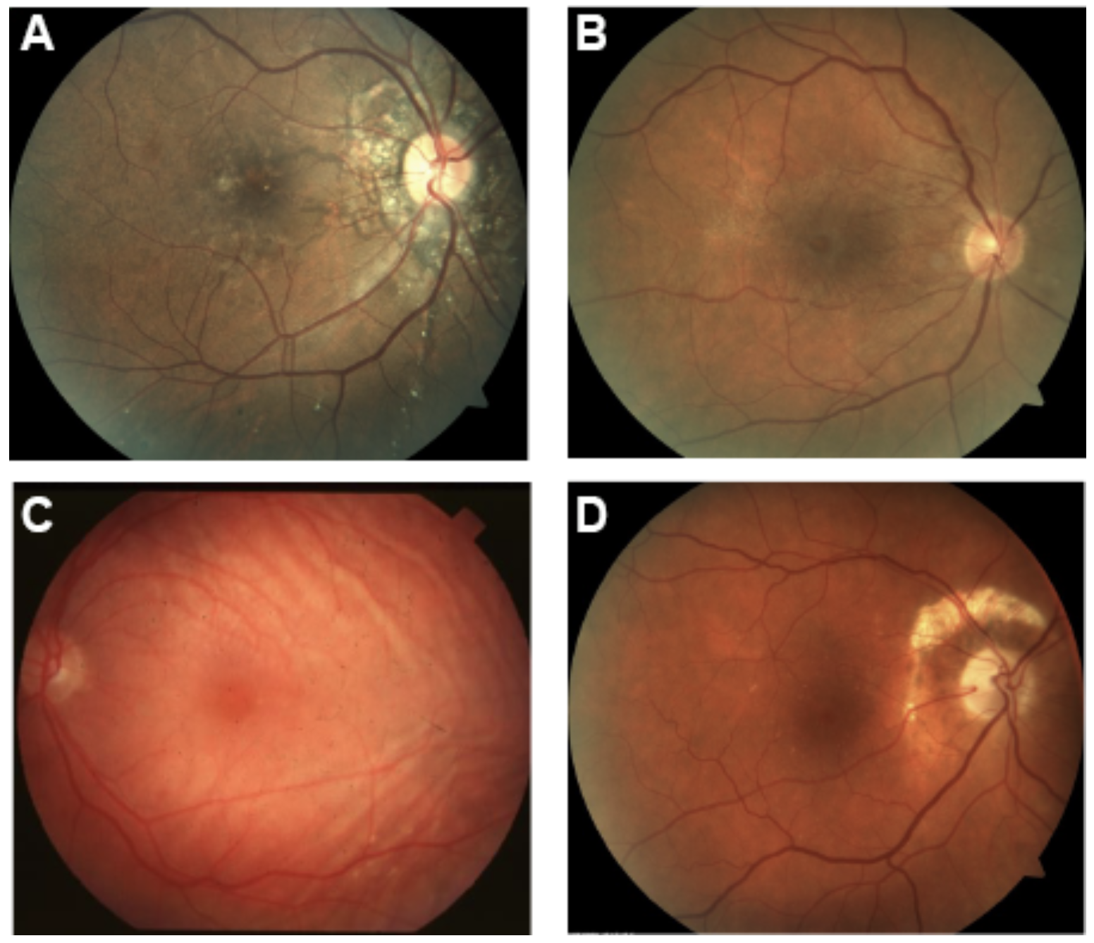
Which of the following images represents angioid streaks?
 |
| Click image to enlarge. |
Answer appears later in newsletter.
JOURNAL ABSTRACTS
OCT Prognostic Biomarkers for Progression to Late Age-related Macular Degeneration: A Systematic Review and Meta-analysis
Imaging biomarkers in intermediate AMD play an important role in helping clinicians recognize the individuals at higher risk for progression to advanced AMD, whether geographic atrophy or neovascular AMD. Over 100 OCT biomarkers have been identified, an exhaustive list of varying significance. This systemic review and meta-analysis sought to determine the biomarkers inferring the greatest risk of progression from early or intermediate AMD to advanced. A total of 1895 research study abstracts were screened with 47 studies ultimately used for the meta-analysis. There were 114 OCT biomarkers across these studies with a calculable risk.
Six biomarkers were found to have high certainty of evidence and greater predictive ability than the traditionally emphasized finding of large drusen. These were abnormalities (attenuation, discontinuity, irregularity etc) in outer retinal bands: the ELM, EZ or IZ. The other three were intraretinal hyperreflective foci, concurrent large drusen and reticular pseudodrusen (also known as subretinal drusenoid deposits) and hyporeflective drusen cores. Intrareflective hyperreflective foci and hyporeflective drusen cores were significantly associated with risk for GA, in particular, whereas EZ abnormalities were more suggestive of progression to neovascular AMD. Other high-risk biomarkers were found but with moderate or low certainty of evidence. With clinical and research importance, this study was able to identify OCT biomarkers that can flag the individuals with AMD at highest risk for progression allowing the potential for better management.
| Trinh M, Cheung R, Duong A, Nivison-Smith L, Ly A. OCT Prognostic Biomarkers for Progression to Late Age-related Macular Degeneration: A Systematic Review and Meta-analysis. Ophthalmol Retina. 2023 Dec 27:S2468-6530(23)00668-1. Epub ahead of print. 1. |
Uveal Melanoma in African Americans: Diagnostic Challenges
Uveal melanoma is very rare in African Americans, with an incidence estimated to be approximately 0.2-0.3 per million people compared to 5.2 per million people in the general US population. The rate of uveal melanoma in Caucasian patients, for example, is believed to be at least 18-fold the rate seen in African Americans. This retrospective record review performed at the Cole Eye Institute in Cleveland identified 12 African American patients that had been diagnosed with uveal melanoma between September of 2004 and December of 2022. This review revealed some interesting findings regarding this group of twelve patients. Five of the tumors were located in the ciliary body / choroid, several were strictly in the choroid, and none were located in the iris. The average diameter of these twelve tumors was 11.5 mm, the average thickness was 6.1 mm, and all of the lesions were brown / dark brown. The large size of these tumors overall may have been related to delayed diagnosis due to a lack of suspicion for uveal melanoma in African Americans. In most instances, primary uveal melanomas exhibit low internal reflectivity on ultrasound while metastatic lesions exhibit high internal reflectivity. Interestingly, four of these tumors exhibited high internal reflectivity on ultrasound even though they were all primary tumors. The authors speculated that this may be due to the fact that very highly pigmented tumors can show high internal reflectivity, and this would be expected in African American patients. Therefore, high versus low internal reflectivity on ultrasound may not be reliable in differentiating factors between primary and metastatic lesions in African American patients. Ultimately, six of the twelve eyes underwent plaque radiation therapy and six were enucleated.
| Yeşiltaş YS, Oakey Z, Wrenn J, et al. Uveal melanoma in African Americans: Diagnostic challenges. Surv Ophthalmol. 2023 Jul 3. Epub ahead of print. |
Surgical Outcomes of Rhegmatogenous Retinal Detachment and Fellow Eye Involvement in Adolescent and Young Adult Patients
The understanding of rhegmatogenous retinal detachment (RRD) is better understood in the adult population as it more commonly occurs in that age demographic from posterior vitreous detachment. But for juvenile and young adults there is far less data on RRD treatment success and fellow eye risk. This retrospective review focused on patients aged 30 and younger who underwent an RRD repair at a retina practice. A total of 109 eyes of 101 patients were included with an average age of 23.85 years (range from 12-30). Myopia (66 eyes), defined as greater than 1D, was the most common associated risk factor, followed by trauma (8 eyes) and previous ocular surgery (7 eyes). The anatomical success rate was high with scleral buckle at 88.7% and scleral buckle with vitrectomy at 89.7% over vitrectomy alone at 75%. As such, the authors stressed the importance of young surgeons acquiring skill with scleral buckle. Of greatest significance for the eye care perspective was the study’s findings of fellow eye risk. Twelve of the patients presented already with bilateral RD. Of those with unilateral findings at presentation, a fellow eye retinal tear or RD developed in 14 of the patients, on average 8 months later. Seventeen patients were followed over 5 years, three of this group went on to have a retinal tear or detachment in the fellow eye. This stresses the importance of carefully monitoring the retina of the fellow eye after development of juvenile/young adult RRD.
Bomdica PR, MacCumber MW, Abdel-Hadi S, Parker M, Minaker S. Surgical Outcomes of Rhegmatogenous Retinal Detachment and Fellow Eye Involvement in Adolescent and Young Adult Patients. Ophthalmol Retina. 2024 Feb;8(2):148-154. |
Risk Factors and Clinical Significance of Prechoroidal Cleft in Eyes with Neovascular Age-related Macular Degeneration in Caucasian Patients
Prechoroidal cleft (PC) is an OCT finding associated with macular neovascularization that is most often found in eyes with age-related macular degeneration (AMD) that have received multiple anti-VEGF injections. It consists of a hyporeflective, optically empty space located between the neovascular tissue and Bruch’s membrane. This retrospective study from Israel evaluated eyes with prechoroidal cleft in neovascular AMD and compared them to eyes with neovascular AMD that did not have prechoroidal cleft. A total of 140 eyes from 140 Caucasian patients were included and were followed for a minimum of 24 months. Of the 140 eyes, 21 (15%) exhibited prechoroidal cleft. All eyes had received multiple anti-VEGF injections starting with Avastin and then switched to other agents only in the case of a poor response to initial therapy. In six eyes with PC, the cleft disappeared at some point during follow-up, but in three of those six eyes it returned. The eyes with PC received significantly more injections (23.4 on average) compared to eyes without PC (18.2). Visual acuity was not significantly different between eyes with PC and those without. Eyes with PC were less likely to have intraretinal fluid than those without but were more likely to have subretinal fluid. In addition, compared to eyes without PC, eyes with PC exhibited greater central subfield foveal thickness, greater maximum retinal thickness, and greater pigment epithelial detachment height. They were also more likely to have multi-layered pigment epithelial detachments. Taken as a whole, these findings may help to predict patients who are most at risk for developing prechoroidal cleft.
Kredi G, Iglicki M, Gomel N, et al. Risk factors and clinical significance of prechoroidal cleft in eyes with neovascular age-related macular degeneration in Caucasian patients. Acta Ophthalmol. 2023 May;101(3):e338-e345. |
Cosmetic Filler-Induced Vascular Occlusion: A Rising Threat Presenting to Emergency Departments
Dermal fillers have emerged as one of the most popular cosmetic treatments in the 21st century. Vascular emergencies from cosmetic filler-induced vascular occlusion are rare occurrences, estimated at 0.01 to 0.05% per treatment. However, in the last two decades, the medical literature has chronicled a tripling of blindness/stroke due to filler-induced vascular occlusions. In patients with skin ischemia involving the ocular region, there is nearly a 50-fold greater risk of vision loss and 10-fold greater risk of stroke compared with those in patients with non-ophthalmic skin injuries. The mechanism of retinal and cerebral ischemic injuries is thought to involve the retrograde flow of a filler embolus toward the proximal ophthalmic and internal carotid arterial systems. Management of filler-induced retinal and cerebral ischemic injuries centers on the rapid delivery of care for vascular occlusions; however, therapeutic success still remains limited. A neuro-ophthalmologic examination should be conducted in all cases, along with an assessment of the oral and nasal cavities.
Hyaluronidase therapy, although highly efficacious in hyaluronic acid–induced skin injuries, has shown minimal benefit in instances of retinal and cerebral ischemic injuries due difficulties in achieving rapid delivery of the enzyme into the orbital and cerebral vasculature. Retrobulbar hyaluronidase therapy, in which hyaluronidase is administered into the posterior orbit, requires expertise and has a high (>80%) rate of failure. Endovascular reperfusion therapy with intra-arterial hyaluronidase performed by interventional radiology along with intra-arterial fibronlytic therapy has recently gained support with some medical professionals.
Soares DJ, Hynes SD, Yi CH, et al. Cosmetic Filler-Induced Vascular Occlusion: A Rising Threat Presenting to Emergency Departments. Ann Emerg Med. 2024 Jan;83(1):59-67. |
Possible Association Between Vaccination Against SARS-CoV-2 and Recurrence of Macular Edema due to Branch Retinal Vein Occlusion: A Case Report
This article details a case report of a patient who developed recurrence of macular edema from a previous branch retinal vein occlusion (BRVO) three days after the administration of a fourth dose of a vaccine against SARS-CoV-2. The patient had received two doses of the SARS-CoV-2 mRNA vaccine BNT162b2 (Pfizer–BioNTech) in 2021 as well as a third dose of the SARS-CoV-2 mRNA-1273 vaccine (Moderna) in March 2022. In January 2023, the patient received a fourth dose of the SARS-CoV-2 mRNA vaccine BNT162b2 (Pfizer–BioNTech). Three days later the patient developed a recurrence of macular edema. The patient was immediately treated with aflibercept and one month after injection, the macular edema resolved and vision returned to baseline.
At present, the association between RVO and vaccination against SARS-CoV-2 remains unclear. The literature has noted that thromboembolic events can occur following vaccination against SARS-CoV-2. Vaccination against SARS CoV 2 may trigger an inflammatory response, microvascular disorders, and hypercoagulability, resulting in the development of a BRVO. Recurrence of ulcerative colitis, glomerulonephritis, multiple sclerosis, nephrotic syndrome, uveitis,and Vogt– Koyanagi–Harada (VKH) disease have also been associated with vaccination against SARS-CoV-2.
| Muto T, Machida S, Imaizumi S, Kamoi K. Possible association between vaccination against SARS-CoV-2 and recurrence of macular edema due to branch retinal vein occlusion: a case report. J Int Med Res. 2023 Nov;51(11). |
IN THE NEWS
PulseSight Therapeutics Launches with Gene Therapy Platform
PulseSight Therapeutics, an ophthalmology focused biotech company, started in February 2024 with a new concept platform of gene therapy. The company uses an electro-transfection system targeting the ciliary muscle for drug delivery of DNA plasmids encoding therapeutic proteins. The technology has been validated in phase 1/2 trial. Their drug candidates include PST-809 and PST-611 targeting wet AMD and geographic atrophy, respectively. Both aiming to reduce treatment burden with need for re-treatment every 6 months.
PulseSight has been financed with seed investment via Pureos Bioventures and ND Capital. They are underway cultivating a Series A financing round to bring programs forward toward clinical proof-of-concept.
Aviceda Progresses into Part 2 of SIGLEC Trial on GA Treatment Candidate AVD-104
Aviceda Therapeutics recently announced positive topline data for part 1 of SIGLEC, a phase 2/3 trial of intravitreal injection of AVD-104 for the treatment of geographic atrophy due to AMD. AVD-104, an intravitreal glycan-coated nanoparticle, boasts a dual mechanism of action that regulates inflammatory cellular and complement pathways. The company reported a positive safety profile, visual improvement, and a reduction in GA lesion growth in part 1. The company has now progressed into part 2, a multi-center, double-masked, randomized, controlled trial to assess safety and efficacy of AVD-104 (either low or high-dose) as compared to complement inhibitor treatment (avacincaptad pegol). Aviceda projects to enroll 300 individuals. The primary endpoint will be the difference in GA growth rate on fundus autofluorescence at 12 months.
Apellis Receives Negative Vote on European Application for Pegcetacoplan
Apellis Pharmaceuticals announced the European Medicines Agency’s Committee for Medicinal Products for Human Use (CHMP) has voted a negative opinion on intravitreal Syfovre (pegcetacoplan); a treatment approved by the FDA for geographic atrophy due to AMD. The EMA released its opinion on January 25th. The EMA website stated “the Agency’s opinion was that the benefits of Syfovre did not outweigh its risks and it recommended refusing marketing authorization”. Apellis has already appealed for re-examination of the marketing authorization application.
OcuTerra Misses the Mark in Phase 2 DR:EAM Trial for DR
Topline results of phase 2 DR:EAM trial were announced by OcuTerra Therapeutics. The clinical trial investigated the eye drop, nesvategrast (OTT166), a selective RGD integrin inhibitor for the treatment of diabetic retinopathy. Although nesvategrast demonstrated a favorable safety profile, the trial did not meet its primary or key secondary endpoints as the data failed to show a statistically significant improvement on the diabetic retinopathy severity scale.
IMAGE QUIZ ANSWER
 |
| Click image to enlarge. |
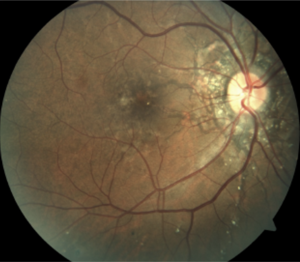
Image A correctly identifies angioid streaks. These represent breaks in Bruch’s membrane. Characteristically, angioid streaks appear bilaterally radiating from around the optic nerve as seen in the fundus image here. Angioid streaks are often associated with systemic disease, most commonly pseudoxanthoma elasticum, Paget’s disease, and sickle cell disease among others. Management consists of observation in particular monitoring for development of choroidal neovascular membrane. Those without a known associated systemic condition should be worked up as appropriate for the most common causes.
B: ERM with pucker
C: choroidal rupture
D: choroidal folds
|
