 |
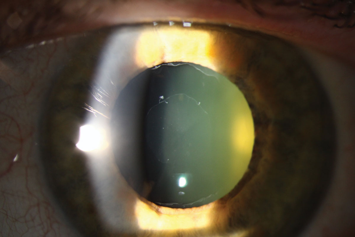
| Mini-Mental State Examination scores were lower in PEX patients with glaucoma, indicative of greater Alzheimer’s severity. Photo: Christine Sindt, OD. Click image to enlarge. |
Editor’s Note: As part of our “Year in Review” retrospective, we’ve selected the top 30 news stories of the year and are re-sharing them as we close out 2023. Follow along as we count down to number 1!
This story was originally published June 14, 2023.
No. 3 biggest news story of 2023:
An age-related disorder characterized by extracellular material accumulation in the anterior eye segment, pseudoexfoliation (PEX) syndrome is not fully understood in its pathogenesis. However, PEX deposition shares features with amyloid aggregation seen in Alzheimer’s disease, making the two possibly interconnected. Just how strongly are they? This was the question researchers from Korea recently posed.
The retrospective design included 48 PEX patients who were age- and sex-matched with the same number of controls. PEX patients were divided into two groups: one consisting of those with glaucoma and the other without. Brain atrophy was the main outcome, which was determined through a visual rating scale from MRI scans and incidence of Alzheimer’s.
Medial temporal atrophy percentage was 56.3% for the PEX group and 35.4% for controls. Global cortical atrophy and parietal atrophy scores were both higher in the PEX group as well. However, there was no difference in brain atrophy between the PEX and PEX glaucoma groups.
A fair portion of the PEX group was diagnosed with dementia (n=16) while only five were diagnosed in the control group. What’s more, PEX glaucoma patients had lower Mini-Mental State Examination scores than those without glaucoma, an indication of more impaired cognitive function in this group. The PEX glaucoma group additionally had fewer mild scores (three out of seven) on the Mini-Mental State Examination, indicating the severity of Alzheimer’s was greater in this group than the PEX group that reported eight out of nine with mild scores.
PEX material formation is not yet understood, but one study found PEX patients with amyloid deposits in the iris arterioles, lens capsule and cornea. PEX material also consists of serine proteinase inhibitors that act as inflammatory substances and regulators of amyloid formation.
Two previous studies also looked into the relationship between PEX and Alzheimer’s with conflicting results. However, the authors of this study point out that both prior ones diagnosed Alzheimer’s from clinical symptoms, while this one used MRI.
There are multiple associations of PEX with amyloid, and amyloid has been identified in the aqueous humor of PEX patients. For these reasons, the authors proposed that “the detection of amyloid in the aqueous humor of patients with PEX predicts an association between PEX and Alzheimer’s. These findings suggest that PEX may be associated with brain atrophy, a common feature of Alzheimer’s disease.”
While PEX is potentially associated with brain atrophy, brain atrophy is a predictive factor of Alzheimer’s pathology; early medial temporal atrophy predicts progression from mild cognitive impairment to Alzheimer’s, and parietal atrophy is seen in preclinical Alzheimer’s patients. As a result, the authors noted that “further investigations are required to determine the relationship between PEX and Alzheimer’s disease with a view to establishing PEX as a predictor of Alzheimer’s.”
Jeong WC, Min JY, Kang TG, Bae H. Association between pseudoexfoliation and Alzheimer’s disease-related brain atrophy. PLoS ONE. June 8, 2023. [Epub ahead of print]. |
