 |
|
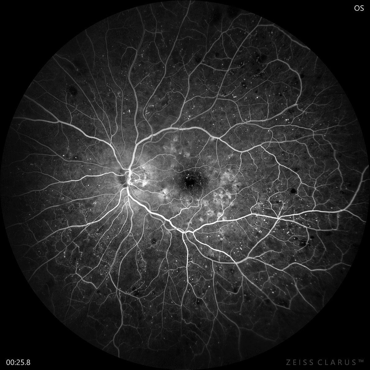
The choroidal vascular density differs depending on where a patient falls on the DR spectrum. Photo: Jay Haynie, OD. Click image to enlarge. |
A recent study aimed to provide a reference for choroidal vascularity by diabetic retinopathy (DR) stage using the choroidal vascular density (CVD) obtained from swept-source OCT (SS-OCT) en face images. The researchers found significant differences in the choroidal vasculature of diabetes patients.
The prospective multicenter study included groups with the following characteristics:
- healthy controls (n=28)
- no DR (n=23)
- nonproliferative DR (NPDR) without diabetic macular edema (DME) (n=50)
- NPDR with DME (n=38)
- proliferative DR (PDR) or any previous treatment with panretinal photocoagulation (n=26)
At the choriocapillaris slab level, the no DR group had a higher CVD and the NPDR with DME and PDR groups had a lower CVD than controls. At the level of the large choroidal vessels, borth the NPDR with DME and PDR groups had a lower CVD than controls.
“This data can play a useful role in choroid assessment in diabetes patients in the future, serve as helpful knowledge to those involved in this activity and contribute to an understanding of the pathology of DR that can be used to identify its mechanism of onset and diagnosis and treatment of the disease,” the study authors wrote in their paper for the Retina journal. “Further research on CVD and diabetic choroidopathy using data obtained from procedures such as OCT angiography and fluorescein may deepen the understanding of DR and diabetic choroidopathy.”
Nakano H, Hasebe H, Murakami K, et al. Choroidal vascular density in diabetic retinopathy assessed with swept-source optical coherence tomography. Retina. October 10, 2022. [Epub ahead of print]. |
