 |
|
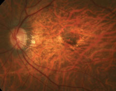
Visual improvement after intravitreal bevacizumab injections for high myopic macular neovascularization is not maintained over time. Photo: Mark Dunbar, OD. Click image to enlarge. |
High myopes find themselves at increased risk of sight-threatening macular neovascularization that requires lifelong observation by eye specialists. Anti-VEGF agents such as bevacizumab have proven effective and safe in treating myopic macular neovascularization and are now the first-line option. Although short-term results look promising, the visual improvement after treatment is of a temporary nature. A recent retrospective study noted that while best-corrected visual acuity (BCVA) seemed to improve significantly at the initial phase, it decreased below baseline after approximately five years.
The analysis included longitudinal clinical data of high myopic macular neovascularization patients treated with intravitreal bevacizumab. High myopia was defined as spherical equivalent (SE) ≤-6.00D or axial length >26.5mm. Active macular neovascularization was confirmed by leakage on fluorescein angiography or OCT-A combined with signs of active macular neovascularization (subretinal fluid) on SD-OCT. Recurrence of myopic macular neovascularization was defined as a reactivation of or new myopic macular neovascularization more than three months after the last injection. The data consisted of 117 eyes of 106 Caucasian patients who were followed from the first injection up to 12 years.
A median of two injections per macular neovascularization episode successfully treated half the patients, while 9% needed more than 10 injections to establish regression of the neovascular lesion.
Mean baseline BCVA of 20/80 significantly improved after the first treatment, to 20/50). At four years (n=86 eyes), BCVA was no longer significantly better than baseline and continued to deteriorate to 20/125 at 10 years (n=27). Of the 27 eyes who reached 10 years of follow-up, 53% developed macular neovascularization-related chorioretinal atrophy. The cumulative incidence of recurrent myopic macular neovascularization was 34% at two years and 59% at five years. BCVA decrease in eyes with or without recurrent macular neovascularization was similar. Patchy chorioretinal atrophy (hazard ratio: 3.0) and subfoveal macular neovascularizations (hazard ratio: 2.5) were significantly associated with recurrent vessel growth.
Eyes with subfoveal macular neovascularization were more likely to develop recurrent lesions. Other factors including age, SE and macular neovascularization size were not significant predictors for macular neovascularization recurrence in this study.
“Lifelong treatment is not necessary for most myopic macular neovascularization: one to 10 injections were enough to cause regression of the macular neovascularization in 91% of cases,” the researchers wrote in their paper. “One out of two eyes developed recurrent myopic macular neovascularization over time, but recurrences did not worsen the course of the BCVA.”
Nevertheless, they noted that large prospective studies using multimodal imaging are needed to build on these findings and optimize treatment and visual outcomes.
Ravenstijn M, Klaver CCW, Yzer S. Long-term treatment outcomes after bevacizumab therapy for macular neovascularization in Caucasian patients with high myopia. Retina. November 16, 2022. [Epub ahead of print]. |
