 |
|
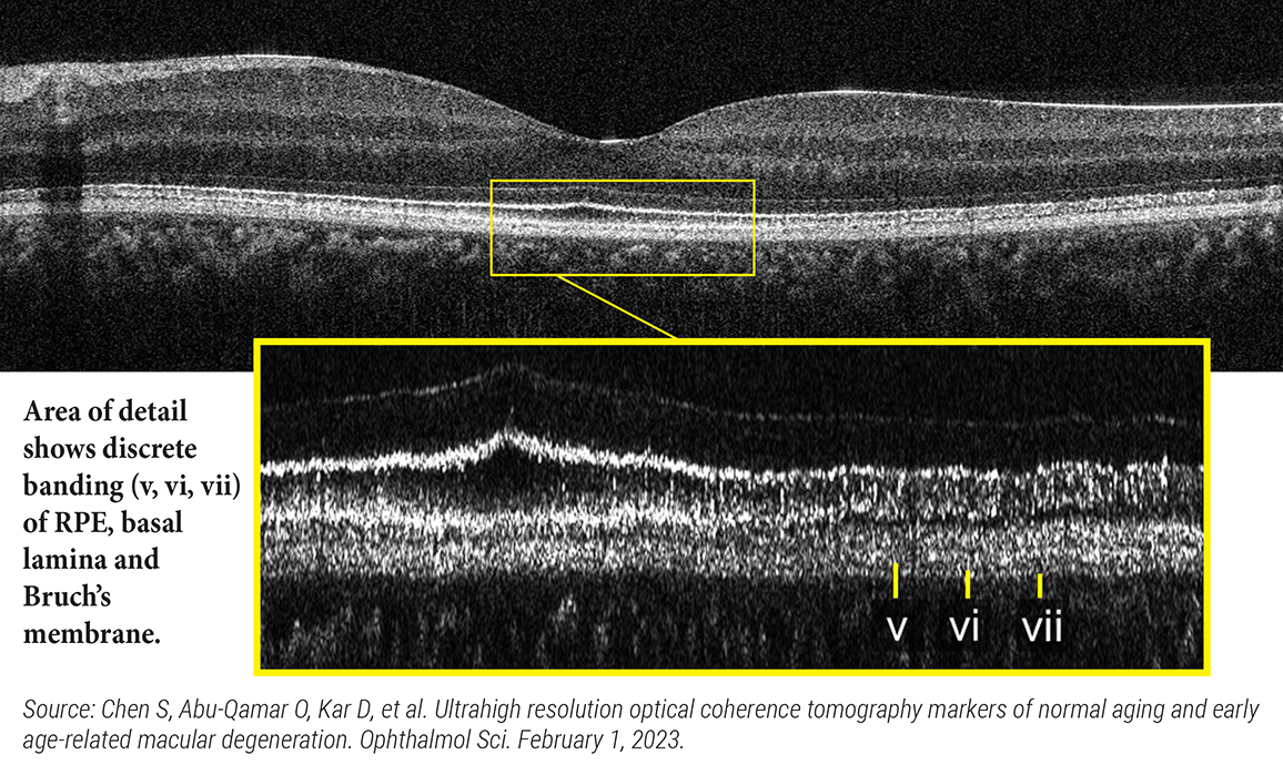
Ultrahigh resolution SD-OCT can enable in vivo and longitudinal assessment of key AMD pathological features, which are typically accessible only ex vivo. In this scan, from a healthy 29-year-old Asian female, the RPE/basal lamina/Bruch’s split is evident. Hyperreflective bands #v and #vii, as well as the hyporeflective band #vi, are resolved across almost the entire field. Photo: Ophthalmology Science [www.ophthalmologyscience.org/article/S2666-9145(23)00009-X/fulltext]; has been enlarged and cropped.). Click image to enlarge. |
Age-related macular degeneration (AMD) affects multiple structures in the retina, particularly the retinal pigment epithelium (RPE), its basal lamina and Bruch’s membrane. The development of markers for disease requires differentiating between normal aging vs. AMD pathology. Using a prototype ultrahigh resolution SD-OCT instrument to image normal adult participants and AMD patients, researchers found that the RPE/basal lamina/Bruch’s membrane complex, seen in commercial OCT as a single hyper-reflective band, can be further resolved as three distinct bands in healthy young adult eyes and in eyes with early AMD. Their recent study demonstrated that ultrahigh resolution SD-OCT is a promising modality that can reveal outer retinal alterations associated with normal aging and early AMD pathology.
Clinical ultrahigh res SD-OCT scans were performed using a high-density protocol on 53 dry AMD eyes from 39 patients and 63 normal eyes from 39 participants. Three trained readers evaluated and labeled outer retinal morphological features, including the appearance of a hyporeflective split within the RPE-RPE basal lamina-Bruch’s membrane complex on ultrahigh resolution B-scans. A semi-automatic segmentation algorithm measured the thickness of the RPE-RPE basal lamina-Bruch’s membrane complex split/hyporeflective band.
In young normal eyes, ultrahigh resolution SD-OCT consistently revealed an RPE-RPE basal lamina-Bruch’s membrane complex split/hyporeflective band. Its visibility and thickness were reduced in older eyes. However, the split/hyporeflective band was again visible in early AMD eyes. Both qualitative reading and quantitative thickness measurements showed significantly elevated visibility and thickness of the RPE-RPE basal lamina-Bruch’s membrane complex split/hyporeflective band in early AMD eyes compared with age-matched controls.
“The hyporeflective band is believed to have a different origin in early AMD than in young normal eyes,” the researchers wrote in their paper. “With evidence from transmission electron microscopy and known AMD pathophysiology, we hypothesize that the hyporeflective band corresponds to the accumulation of basal laminar deposit in early AMD eyes.”
The team noted that longitudinal studies, especially on older eyes that develop AMD and early AMD eyes that progress to intermediate AMD, are crucial to test the diagnostic and predictive performance of imaging markers, while further verifying their relationship with pathogenesis.
“Ultrahigh resolution SD-OCT can be used to investigate physiological aging as well as early AMD pathology in clinical imaging studies,” their paper concluded. “Developing quantifiable markers associated with disease pathogenesis and progression can facilitate drug discovery, as well as reduce clinical trial times.”
Chen S, Abu-Qamar O, Kar D, et al. Ultrahigh resolution optical coherence tomography markers of normal aging and early age-related macular degeneration. Ophthalmol Sci. February 1, 2023. [Epub ahead of print]. |
