 |
| Although all imaging systems performed well in this study, the authors caution that shifting from one platform to another could change the patient’s score on the Diabetic Retinopathy Severity Scale. Photo: Julie Torbit, OD. Click image to enlarge. |
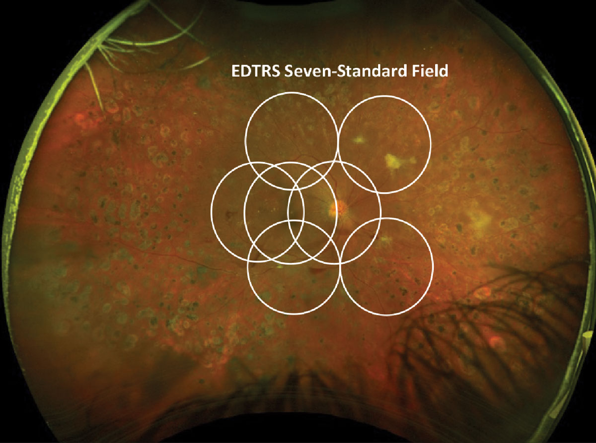
The field of retinal imaging has advanced significantly in recent years, boosting the reliability and accuracy of diagnosis in many cases. Along with this advancement, current literature should be updated on how different ultra-widefield imaging compares. This task was taken by a team of researchers who compared diabetic retinopathy (DR) severity levels assessed from seven standard-field (7F) stereoscopic color photographs on a 35° fundus camera to both Clarus and Optos ultra-widefield color images.
Included in the study were 97 eyes (50 patients) that were imaged with the Topcon 35° fundus camera, Clarus and Optos. The DR Severity Scale (DRSS) was determined within the 7F area of each image set using the Early Treatment Diabetic Retinopathy Study scale. The researchers found agreement within one step of ETDRS levels between standard 7-field imaging and Clarus 7F to be 90.1%, while it was 85.9% with Optos. Agreement within one step between standard 7-field imaging and Clarus global was 88.9% of eyes, while Optos global was 85.7%. Agreement between Clarus and Optos global DR level within one step was 89.1%, and intergrader agreement of the 7F ETDRS level was 96.0% for standard 7-field imaging, 98% for Clarus and 95.5% for Optos.
With this information, they study researchers were able to conclude moderate to substantial agreement between all three devices spanning the full spectrum of DR levels. Equivalency was considered at high agreement rate between DR level within one step between modalities and two-step agreement is accepted for clinical trial outcomes. Here, one-step agreement was greater than 85% and two-step agreement between all three imaging types was more than 95%, supporting “the notion that both Clarus and Optos are equivalent to standard 7-field imaging for obtaining 7F ETDRS levels in DR clinical trials,” the researchers wrote in their paper for Ophthalmology Science.
This research is significant because although there has been literature outlining Optos in comparison to standard 7-field imaging, the same attention has not been given to agreement of Clarus and Optos systems in assessing ETDRS levels.
The authors point out that NPDR vs. PDR distinction is important, since PDR carries much higher risk of vision loss and requires more aggressive intervention. They do note that NPDR/PDR discrepancy occurred in both directions within every comparison pair, with the shift ranging from 2% to 7%.
Occurrence of PDR outside the 7-field region was also measured. PDR was seen compared to standard 7F in global DR assessment in 5.6% of Clarus images and 8.2% of Optos images. Observed shift from NPDR to PDR status was seen from 7 fields to global ETDRS levels in 4% of Claus images and 2.7% of Optos ones. This conflicts with earlier research that indicated the region beyond the 7F does not significantly affect the DRSS.
For clinical application, the authors advise that “if a facility has access to multiple types of imaging systems, switching between different devices could potentially result in a patient’s DRSS level changing due to variations in the imaging system; this could affect the accuracy of monitoring disease progression. Therefore, it is necessary for clinicians to be aware of the relative features of each camera to ensure reliable tracking of a patient’s DR severity.”
Duncan N, Barrett N, Schildroth K, et al. Comparison of standard 7-field, Clarus and Optos ultra-widefield imaging systems for diabetic retinopathy (COCO Study). Ophthalmol Sci. November 11, 2023. [Epub ahead of print]. |
