 |
|
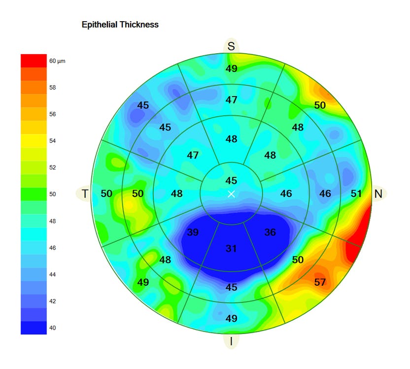
Ectatic diseases such as keratoconus may manifest in the epithelium before other evidence appears. Photo: Carl Zeiss Meditec. Click image to enlarge. |
While the early stages of keratoconus (KCN) can be difficult to identify, one new study offers a promising data that may improve diagnostic accuracy. The research centered around testing whether the OCT metric of corneal epithelial pattern standard deviation (PSD) could distinguish between normal eyes and those with subtle ectatic changes.
The retrospective study compared four groups: healthy, unaffected eyes (n=105); patients with bilateral keratoconus (n=86); very ectatic eyes without any history of surgical treatment (n=11); and fellow eyes of the ectatic cases but with normal topographies (n=61). OCT was used to scan corneas and their epithelial thickness maps were analyzed.
Results showed significant differences in epithelial PSD across all four groups. The normal group of eyes had lower PSD values than both ectasia groups. Additionally, both the KCN and ectasia groups had higher scores than the unaffected fellow eyes, and ectatic eyes had higher recorded epithelial PSD values, even in the fruste form of the condition.
Based on these findings, the PSD variable was able to accurately distinguish clinical ectasia but was not able to identify the fruste form of the disease by itself. In this study, those comprised of the individuals with very asymmetric keratoconus with another normal tomographic eye.
Despite the corneal topography revealing cases that would have gone unnoticed by a slit-lamp and absence of visual impairment, some normal topographic maps still develop complications, potentially due to epithelial thinning that sometimes accompanies apex steepening in mild corneal ectasia. This fact prompted the researchers to suggest that identifying these types of cases might be aided by identifying focal epithelial thinning.
The authors do note that they did not look at variations in epithelial PSD scores at different stages of keratoconus, prompting them to recommend that future studies should see if there is a correlation between those values and severity of the disease. They also add that epithelial PSD can detect all types of abnormalities, not just keratoconus or ectasia. Thus, it is prudent to keep in mind that the healthy group could very well have had epithelial deformations from other conditions like dry eye or epithelial dystrophies while still displaying normal topographic patterns.
The authors are hopeful, though, about the use of this technique to identify early-stage ectatic disease, perhaps when combined with machine learning tools. “Our next step is to apply artificial intelligence methods,” they wrote in their paper, “to combine epithelial data with the tested tomographic and biomechanical parameters, which might provide a robust and innovative combination of data to enhance our ability to identify very early forms of ectatic disease.”
Salomão MQ, Hofling-Lima AL, Esporcatte LPG, et al. Corneal ectasia detection by epithelium pattern standard deviation from optical coherence tomography. J Catar Refr Surgery. 2022. [Epub ahead of print]. |
