 |
A 54-year-old woman presented to the office with a chief complaint of needing new reading glasses. She had no other ocular issues. She did not report any pain. The patient also denied trauma, systemic disease or allergies of any kind.
Clinical Findings
Her best-corrected entering visual acuities were 20/20 OD and OS at distance and 20/30 at near through her progressive bifocal spectacles. Her external examination was unremarkable, with no evidence of afferent pupillary defect. Goldmann applanation tonometry measured 17mm Hg OU.
Refraction uncovered a stable distance refraction and the need for more add power in the bifocal (the add was properly positioned in the frame beginning at the bottom of the pupil).
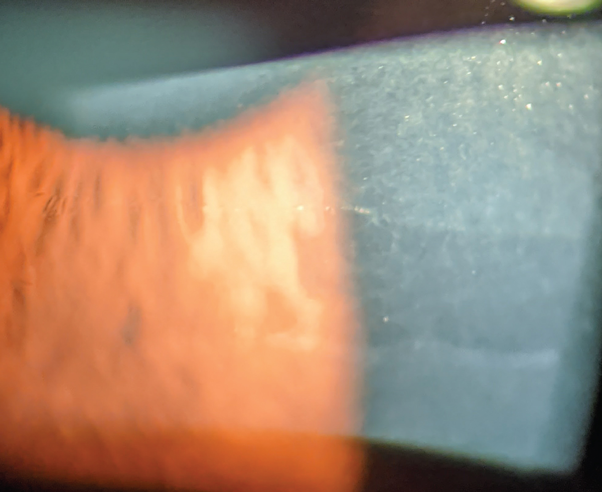
While examining the anterior segment during biomicroscopy, an unexpected pertinent finding was discovered and is demonstrated in the photograph.
 |
|
Her presentation was similar to this one from a patient with the same condition. Photo: Marcus Noyes, OD. Click image to enlarge. |
For More Information
Additional studies might include corneal topography to ensure the corneal surface is regular. Corneal staining with sodium fluorescein dye would permit understanding of the cornea’s overall health status and level of hydration.
What would be your diagnosis in this case? What is the patient’s likely prognosis?
Diagnosis
The diagnosis in this issue is posterior polymorphous dystrophy (PPMD). Corneal dystrophies typically result in gradual bilateral visual changes in eyes with variable symptoms.1,2 The term dystrophia is derived from the prefix dys-, meaning “bad or difficult,” and trephein, meaning “to nourish.” Thus, the meaning implied by the term dystrophy is a malady produced by poor nutrition or metabolism.1 The disease is genetically transmitted by autosomal dominant genes causing aberrant development of the cornea’s Descemet membrane and endothelium.3
PPMD may present unilaterally or bilaterally and is typically discovered within the first decade of life.1-4 Most cases are asymptomatic, with severe loss of function or discomfort leading to corneal transplantation in approximately 25% of affected individuals.5 Posterior polymorphous corneal dystrophy has an estimated prevalence of 1:100,000.6
The disease has the potential to produce corneal opacities (vesicle-like lesions, band lesions and geographic/diffuse opacities), scarring and edema that leads to decreased visual acuity, and the formation of irregular astigmatism—even keratoconus.4,6,7,8 When the disease emerges in individuals of young age, amblyogenic factors can induce meridional and refractive amblyopia.4,6,9,10
The disease is known to share an association with Alport’s syndrome.11,12 This inherited disease is characterized by progressive renal failure, hearing loss and ocular abnormalities.12 Inheritance is X-linked (85%) or autosomal recessive (15%).10 While Alport’s syndrome primarily thought of by many as a male disease, twice as many women are affected by X-linked transmissions.12
Treatment of PPMD is intuitive and will include some combination of the following in appropriate circumstances:1-12
- monitoring and patient education for asymptomatic cases
- artificials tears and lubricants for cases needing moisture
- hypertonic ointments and drops for cases with edema
- collagen crosslinking for cases experiencing corneal warpage or keratoconus formation
- contact lens options ranging from soft, rigid to scleral for cases requiring refractive therapy
- vision training for amblyopia rehabilitation
- surgical options (e.g., Descemet’s stripping endothelial keratoplasty, penetrating keratoplasty) for severe advanced unresponsive cases
Clinical Course
We treated this patient supportively with artificial tear drops and ointments at bedtime. Her lack of signs and symptoms dictated a conservative approach. We did educate her to the presence of the disease process, making her aware that treatments were available to her if new signs or symptoms developed.
Dr. Gurwood is a professor of clinical sciences at The Eye Institute of the Pennsylvania College of Optometry at Salus University. He is a co-chief of Primary Care Suite 3. He is attending medical staff in the department of ophthalmology at Albert Einstein Medical Center, Philadelphia. He has no financial interests to disclose.
|
