 |
As optometry continues to lobby for expanded scope of practice, a lot of attention is focused on the use of lasers. An area equally important for most optometrists to incorporate into their practice is the injection of corticosteroids into chalazia. The procedure is straightforward and provides a non-surgical option to management. Surgical incision and curettage of a chalazion has a high success rate (upwards of 92% after a single procedure) but is also an invasive technique requiring the injection of an anesthetic, clamping of the lesion, incision and subsequent curettage.1
An alternative that is very attractive to many patients is an injection of triamcinolone acetonide into the lesion. Several studies have demonstrated the success of intralesional steroid injections for chalazia with a success rate in some studies over 90%, depending on the concentration of the steroid injected and number of injections.2
 |
|
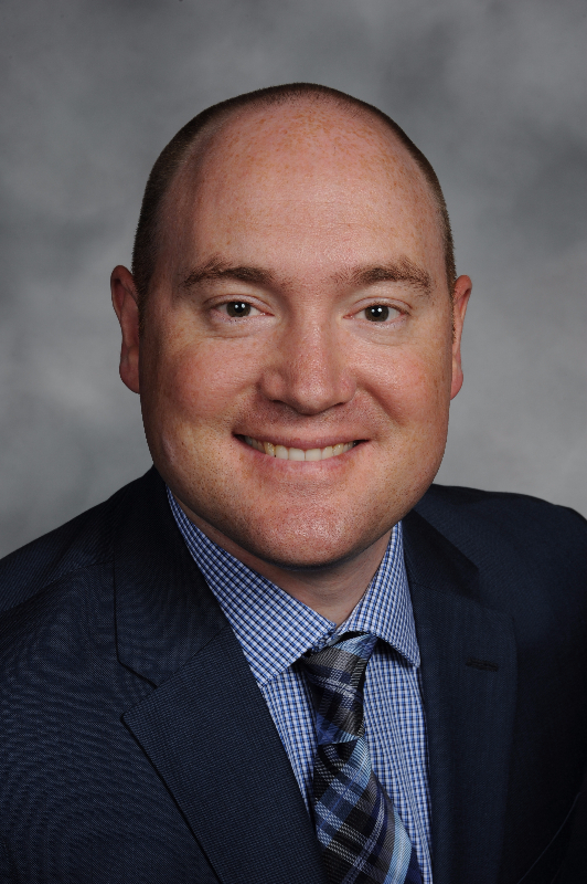
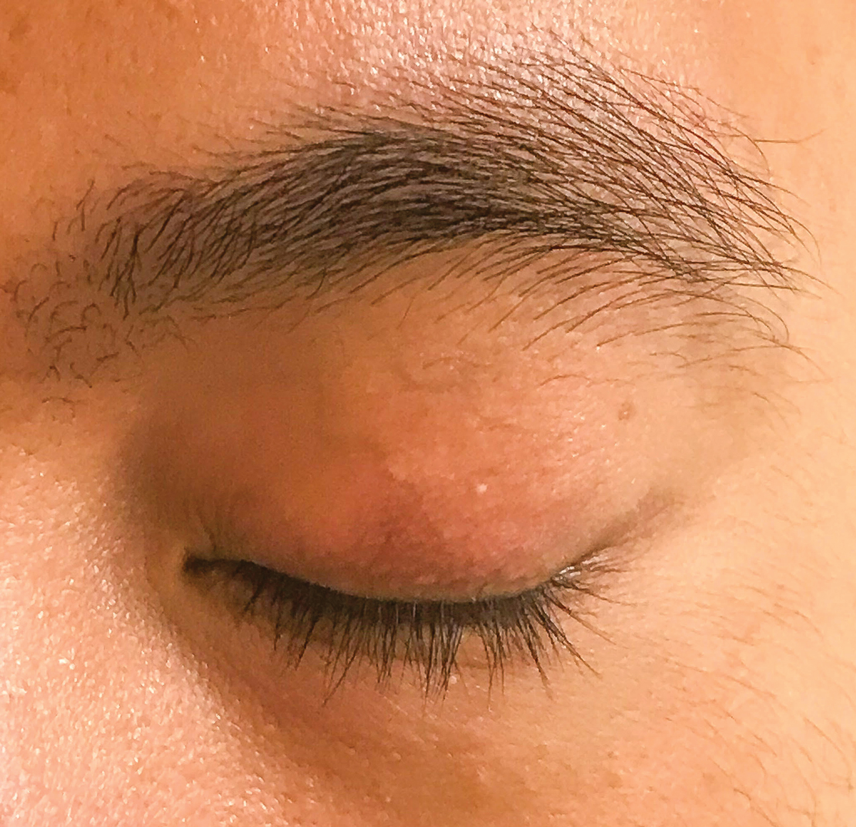
Fig. 1. Chalazion of the left upper lid that has been present for one month. Click image to enlarge. |
Background and Mechanism
The use of depot steroid of triamcinolone acetonide has been widely used in many ophthalmic diseases and procedures such as intralesional injections of granulomas and treatment of periocular scars. It has anti-inflammatory, anti-VEGF and antifibrotic properties.3 Chalazia are composed of corticosteroid-sensitive inflammatory cells. These inflammatory cells cause exudation, which can compress the lymphatic vessels and lead to an alteration in lymphatic vessels that results in granuloma formulation in the lids. The presumed action of depot corticosteroid is suppressing additional release of inflammatory cells and reducing exudation of plasma fluids. The inhibition of further production of inflammatory cells and reduction of plasma fluid exudation can lessen the compressive effect and facilitate the lymphatic absorption of the chalazion’s contents.4
Triamcinolone acetonide is the typical corticosteroid used in intralesional injections. A single dose of intralesional injection of higher concentration of triamcinolone acetonide at 40mg/mL is more effective than at 10mg/mL; however, there is also an increased chance of potential complications.5 There are two main routes of injection—transcutaneous or transconjunctival—with or without topical or injected anesthesia.
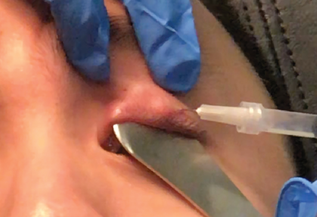 |
Fig. 2. Injection of triamcinolone acetonide into and around the chalazion showing proper angle of the needle. Click image to enlarge. |
Transcutaneous injection tends to be the most popular as there is less pain for the patient and is often the easiest and most efficient. The transconjunctival route is preferred for those patients with whom there is a larger concern for skin depigmentation at the injection site.1 The most common adverse reaction to this treatment is skin depigmentation (particularly in darkly pigmented patients), but using a transconjunctival approach to injection reduces this risk. Triamcinolone acetonide is commonly used for many skin lesions, and skin hypopigmentation has been reported in 1.3% to 6% of cases.6
The advantages of intralesional injection of this steroid are many: it is a simple outpatient procedure typically with little to no bleeding, anesthesia injection is not required, multiple chalazia can be treated at a time, there is no post-procedure scarring and it is useful in children and anxious patients. Potential complications that have been reported after injection of a chalazion include hypopigmentation or depigmentation of lid skin, yellow or white deposits in the lid skin, subconjunctival eyelid fat atrophy, steroid-induced glaucoma, subcapsular cataract and second injection. Reported complications from the injection process include possible corneal perforation with traumatic cataract by injection needle, as well as microembolism of retinal and choroidal vasculature that can lead to infarction.5
Procedural Technique
The equipment needed typically includes:
- Topical anesthetic drops (i.e., proparacaine).
- Alcohol pads for cleaning the top of the medication bottle and for the patient’s lid.
- A 1cc or 3cc syringe.
- An 18-gauge needle for medication draw up.
- A 25- to 27-gauge needle for the injection.
- 10mg/mL or 40mg/mL triamcinolone acetonide (the two of us exclusively use 40mg/mL).
- Gauze for any potential bleeding and putting pressure on lid after injection.
- A sharps container.
- A betadine swabstick for additional disinfection of the lid (optional).
- A Jaeger plate for protection of the globe during the injection (optional).
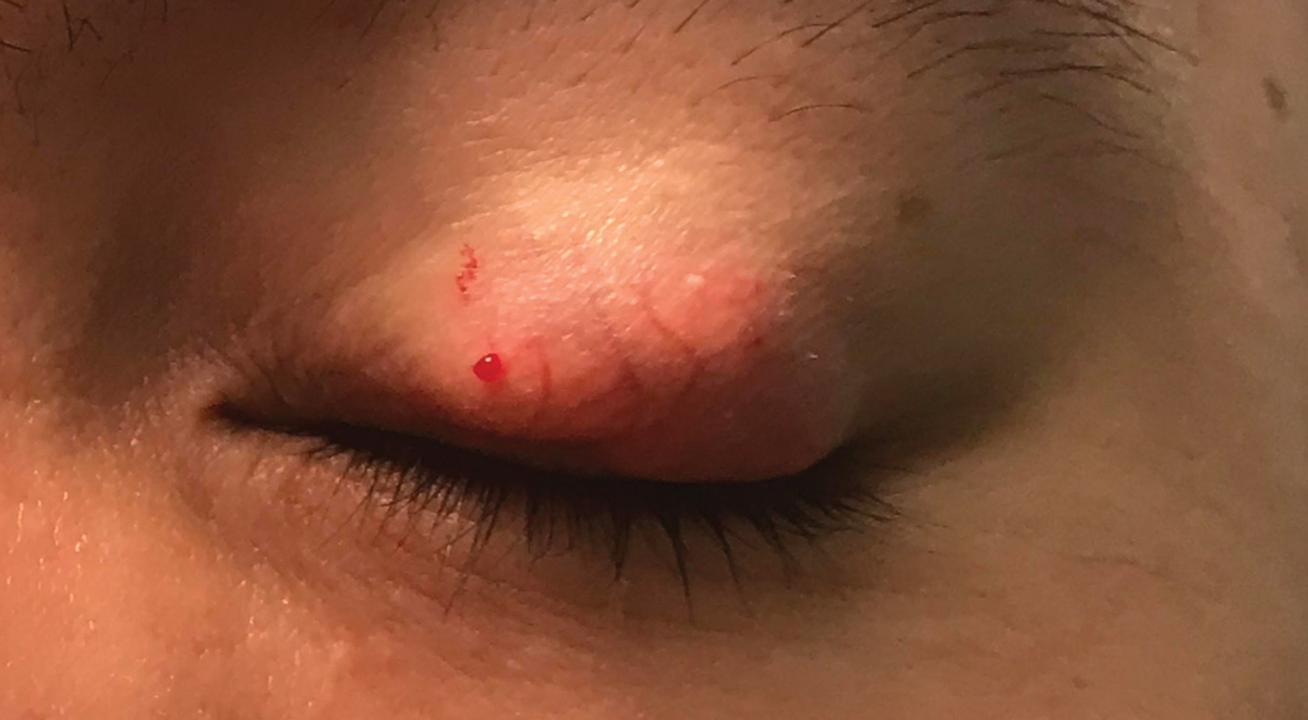 |
| Fig. 3. Patient has received an injection of triamcinolone 40mg/mL from both the temporal and nasal aspect of the chalazion. Click image to enlarge. |
Prior to the procedure, educate the patient about the options available to them, which include leaving the lesion alone (as this is a cosmetic reason for treatment), intense pulsed light therapy, intralesional injection or incision and curettage. Make them aware about possible complications.
Skin depigmentation is likely the most concerning side effect of the injection. I have not seen any depigmentation in Asian or Latinx patients. A patient of African American descent, darkly pigmented skin or one that is concerned about skin depigmentation is often recommended a transconjunctival injection or other potential treatment options. Have the patient fill out and sign a patient consent form that describes the procedure in layman’s terms and potential side effects and have both the patient and the practitioner sign it.
 |
|
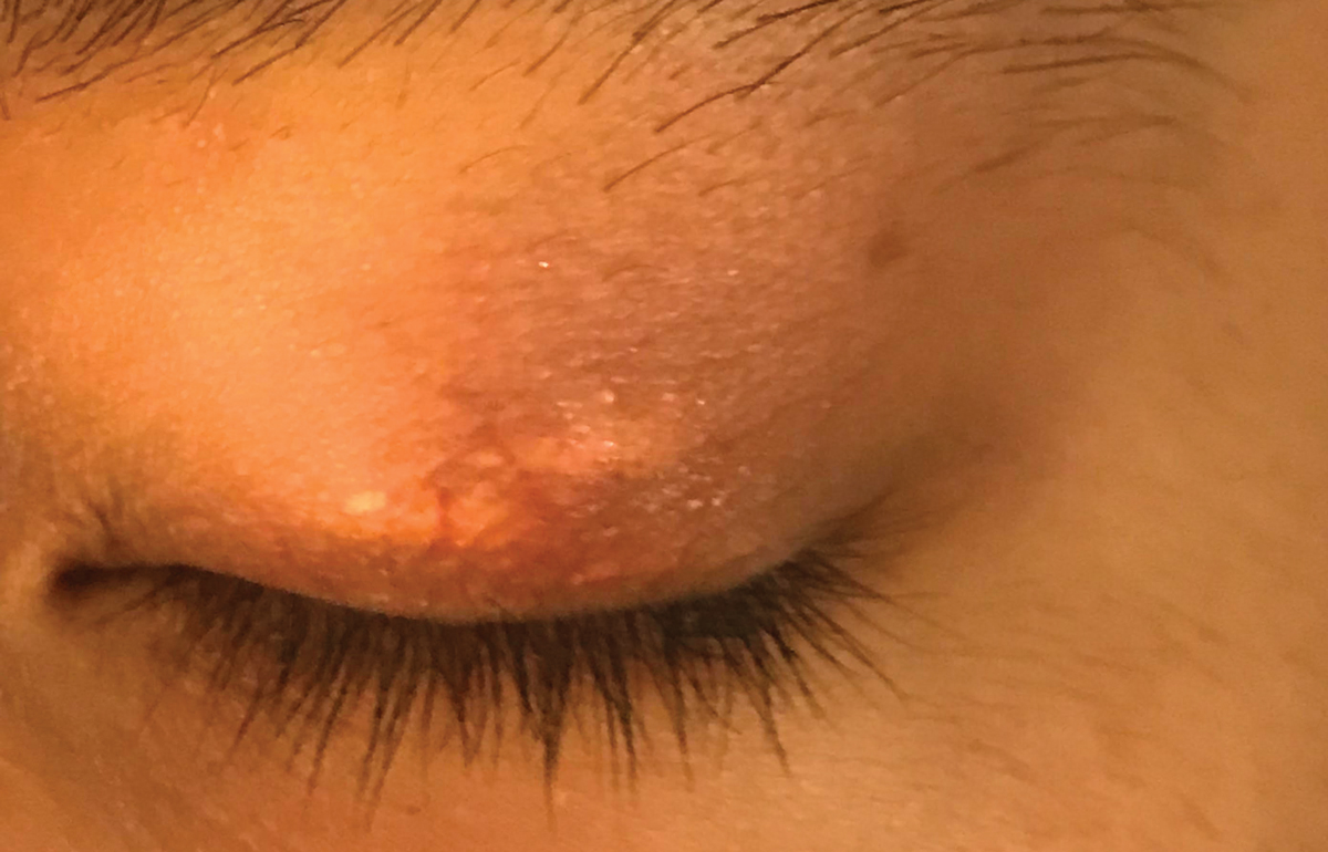
Fig. 4. Skin deposits of steroid after the injection. Let the patient know this will happen and is not skin depigmentation. Click image to enlarge. |
Figure 1 depicts a patient with a chalazion of the left upper lid that has been there for approximately one month. The patient agreed to proceed with a 0.3cc triamcinolone acetonide 40mg/mL injection. A drop of anesthetic was instilled into each eye, and the left upper lid was cleaned with an alcohol wipe. The patient was reclined in the exam chair so that we could approach them from behind to perform the injection. Most injections are done at an angle, but the approach with a chalazion injection is completely parallel to skin (Figure 2). If the lesion is approached at an angle, it is more likely for the needle to go through the lid and possibly hit the globe. There is the option to use a Jaeger plate, which is applied under the lid to protect the globe; however, the plate stretches the skin and can make localization of the chalazion more challenging.
At first impression, you think you would want to inject directly into the chalazion; however, it is difficult to inject into a solid lesion. It is better to start your injection into the skin in front of the lesion and then push the needle forward until it pierces the lesion. As you begin to inject the steroid, you will notice that it is challenging to push it out of the needle; that is because you are in a solid lesion. Don’t push harder and end up moving the needle forward through the lesion. As you continue to try and push steroid out of the needle, you are going to slowly pull out of the lesion, and you will see the skin puff up, indicating that you are injecting steroid into the skin that surrounds the lesion. If the lesion is larger, you can inject the other side of the lesion in the same way by approaching it completely parallel to the skin and piercing the skin outside of the lesion first. Figure 3 shows the lid after the injection of steroid from both the temporal and nasal side.
Post-Procedure and Follow-Up
Tell the patient to leave the lesion alone for several days, i.e., to not push or massage the area. It is important to try and not force the steroid away from the lesion. The steroid is in suspension, and the liquid aspect of the steroid gets absorbed rapidly, leaving behind a depot or deposit of steroid. Figure 4 depicts the deposits of steroid in the injection site. These deposits are not skin depigmentation and may last from four to six weeks after the injection. After three to four days, the patient can begin their warm compresses and gentle lid massage again.
 |
|
Fig. 5. Resolution of the chalazion four to five weeks post-injection. Click image to enlarge. |
Follow-up with the patient typically in two weeks to see how they are progressing. If at two weeks, the lesion looks reduced and there are still steroid deposits in the skin, the patient is told to continue the warm compresses and lid massage and to return if the lesion doesn’t completely resolve. Figure 5 shows the patient four weeks after their steroid injection with complete resolution of the chalazion.
Conversely, if at the initial two- to three-week follow-up the lesion does not look like it has reduced and there are no steroid deposits, give the patient the option of another injection. Alternatively, they can continue to do the warm compresses and massage and return in another two weeks for an injection if there is no further resolution. Keep educating patients on how to prevent further development of chalazia with good lid hygiene practices.
Be sure to document all aspects of the procedure and patient education in both the assessment and plan area, as well as in the narrative of the procedure order (CPT 11900: injection, intralesional; up to and including seven lesions). You will also need to document the amount and concentration of triamcinolone injected, the lot number and the expiration date.
Incorporating steroid injections into your practice is a relatively straightforward process with limited specialized equipment and will provide a much-needed treatment option for your patients. The first injection is the scariest! However, after that one you will feel comfortable that this is a procedure that optometrists can readily offer our patients.
Dr. Lonsberry is a professor of optometry at Pacific University College of Optometry.
1. Aycinena AR, Achiron A, Paul M, Burgansky-Eliash Z. Incision and curettage vs. steroid injection for the treatment of chalazia: a meta-analysis. Ophthalmic Plast Reconstr Surg. 2016;32(3):220-4. 2. Mohan K, Dhir SP, Munjal VP, Jain IS. The use of intralesional steroids in the treatment of chalazion. Ann Ophthalmol. 1986;18(4):158-60. 3. Bartlett JD, Jaanus SD. Clinical Ocular Pharmacology. 5th ed. Butterworth-Heinemann/Elsevier; 2008. 4. Aglianó M, Lorenzoni P, Volpi N, et al. Lymphatic vessels in human eyelids: an immunohistological study in dermatochalasis and chalazion. Lymphology. 2008;41(1):29-39. 5. Lee JW, Yau GS, Wong MY, Yuen CY. A comparison of intralesional triamcinolone acetonide injection for primary chalazion in children and adults. ScientificWorldJournal. 2014;2014:413729. 6. Martins N, Polido-Pereira J, Caneira M, Fonseca JE. Treatment of persistent cutaneous atrophy after corticosteroid injection with fat graft. Reumatol Clin (Engl Ed). 2019;15(6):e122-4. |
