 |
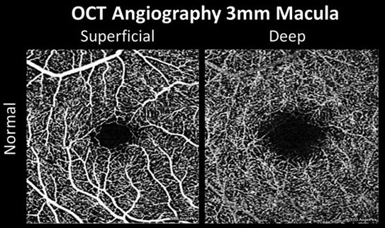
| New research may be laying the groundwork for clinical application of OCTA in amblyopia diagnosis through analysis of superficial and deep vessels. Photo: Carolyn Majcher, OD. Click image to enlarge. |
The emergence of a new technology always brings with it new techniques and insights, and the rise of OCT angiography has been an exemplary case of that, even if some of the findings aren’t ready for mainstream use just yet. A new literature review appearing in Ophthalmic Research examined the macular microvasculature in patients with amblyopia to see if OCTA might be able to one day play a role there, with encouraging results.
In analyzing 17 studies, the researchers found that on 3x3mm2 scans, amblyopic eyes displayed lower whole vessel density of the superficial capillary plexus (SCP) when compared with controls. However, differences became clearer on 6x6mm2 scans, with amblyopic eyes displaying lower perifoveal vessel density of superficial and deep capillary plexus. Both scan types showed whole parafoveal vessel density of deep capillary plexus (DCP) and parafoveal-SCPVD to be lower.
Between amblyopic eyes and fellow eyes, the only difference in all quadrants was that of parafoveal and perifoveal-SCP density and the foveal-DCP density. As well, superficial vessel density was higher in fellow eyes compared to normal eyes in all quadrants but the fovea, while deep capillary plexus vessel density was higher in all quadrants except the parafoveal.
The study researchers reveal primary findings from similar studies, pointing out inconsistencies. One saw no differences in superficial vessels, deep vessels and foveal minimum thickness between the amblyopic and its fellow eye. Another saw no difference with amblyopic eyes, fellow eyes of those with unilateral amblyopia and controls in foveal avascular area, superficial vessel density and deep vessel density.
Meanwhile, another saw lower vessel density of deep capillary plexus in amblyopia patients. An additional study saw both superficial and deep structures to be lower in amblyopic eyes in all quadrants except the fovea with the foveal avascular area smaller in the amblyopic group as well, although the difference was not significant.
Compared with these differences, the study researchers highlight their discovery of differences revealed in normal control eyes compared with contralateral, non-amblyopic eyes in 6x6mm2 scans. Although most indicators were statistically significant, the 6x6mm2 scan more easily detected these observations.
As such, the study authors believe that “this indicates that the development of amblyopia not only affects the macular structure of the affected eye, but also the contralateral non-amblyopic eye, which reminds us that we should pay attention to the changes in both eyes in the diagnosis and treatment of amblyopia.”
Following this thought, the study authors believe that, consequently, OCTA may be used as a diagnostic tool to identify and monitor patients with amblyopia. “However, additional investigations are needed to figure out whether OCTA can be a diagnostic aid in different types of amblyopia via prospective studies with a larger sample size.”
Feng F, Yu Y, Jingyue Z, et al. Evaluation of retinal microvasculature features in patients with amblyopia based on optical coherence tomography angiography: a systematic review and meta-analysis. Ophthalmic Res. March 14, 2023. [Epub ahead of print]. |
