History

A 69-year-old black female presented with a chief complaint of metamorphopsia (O.S.>O.D.). According to her previous optometrist, her ocular history was remarkable for “growing blood vessels and bleeding” in her left eye. Her systemic history indicated that she was being treated for both hypertension and arthritis. The patient reported good compliance with her medications. She denied having any allergies.
Diagnostic Data

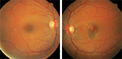
This 69-year-old black female with a history of hypertension presented with a chief complaint of metamorphopsia (O.D. left, O.S. right). What do you notice?
Her best-corrected visual acuity measured 20/25 O.D. and 20/30 O.S. Pupils were equal and round, with no evidence of afferent defect. Extraocular muscle movements were full and unrestricted. Confrontation visual field testing uncovered an irregularity located superior temporal to fixation in her left eye. Accordingly, formal Amsler grid testing confirmed an area of metamorphopsia located superior temporal to fixation in her left eye.
Her intraocular pressure measured 13mm Hg O.U. Slit lamp examination revealed trace nuclear sclerotic cataracts in both eyes. Figures 1 and 2 illustrate the pertinent retinal findings.
Discussion
Other useful tests include automated visual fields, photodocumentation, optical coherence tomography with fluorescein angiography (FA) and indocyanine green (ICG) angiography completed at the retinology appointment.1-4 In such cases, you should also issue an Amsler grid to the patient so he or she may monitor the irregularity for progression at home while waiting to see the retinal specialist..
This patient has idiopathic polypoidal choroidal vasculopathy (IPCV) in her left eye. IPCV has been described as “posterior uveal bleeding syndrome” by Robert C. Kleiner, M.D., and associates and as “multiple recurrent retinal pigment epithelial detachments” by R.M. Stern, M.D.1,2 Then in 1997, Lawrence A. Yannuzzi, M.D., and associates first coined the term IPCV, which was more appropriate due to the condition’s unknown etiology and pathogenesis.3
Typically, IPCV affects hypertensive, black females between the ages of 50 and 65.3-9 Other studies have shown that Asian patients tend to have this condition less frequently than black patients, but more frequently than white patients.3,4 The condition can present either unilaterally or bilaterally with recurrent hemorrhagic and exudative detachments of the retinal pigment epithelium (RPE) in the absence of drusen, degenerative myopia, ocular inflammation or lacquer cracks.3,4
The typical fundus findings in patients with IPCV include reddish-orange juxtapapillary subretinal masses.3-9 These subretinal masses represent abnormal polypoidal branching of the choroidal vasculature.3-9 The polypoidal lesions are associated with multiple recurrent serous detachments of the RPE.3-5 Their size is dependent upon the diameter of the vessels in which they originate.3 Their progression in size is likely caused by simple proliferation and hypertrophy of the vascular components.3-9
The pathogenesis of IPCV is still unknown; however, remnants of old ocular inflammation, a choroidal tumor or a vascular anomaly have all been hypothesized as possible causes.3
One case study indicated that hypertension plays a significant role in the pathogenesis of IPCV.5 The study also suggests that stress placed on the inner choroidal vascular system in hypertensive patients may cause vascular remodeling with focal constrictions and aneurysmal dilations.3 It is believed that these aneurysmal dilations protrude anteriorly through an already damaged Bruch’s membrane.5 This process ultimately leads to a serous detachment of the RPE and vitreous hemorrhage. Choroidal neovascular membrane leakage or polypoidal dilations may lead to metamorphopsia and vision loss.5
Differential diagnoses include wet AMD, choroidal hemangioma, choroidal metastasis, choroidal osteoma, posterior scleritis, retinal arterial macroaneurysm, birdshot choroidopathy, acute posterior multifocal placoid pigment epitheliopathy, panuveitis, multifocal choroiditis and Vogt-Koyanagi-Harada disease.3,6
Treatment of IPCV includes a retinology referral, semiannual dilated fundus examination and home Amsler grid monitoring. If progression is noted or visual acuity is lost, an FA or ICG angiogram is indicated to assess the areas of leakage. If the polypoidal structures begin to encroach into the macula, focal laser photocoagulation (with parameters similar to those used in the treatment of choroidal neovascularization in AMD) may be indicated depending upon the location and extent of the vascular irregularities.3 Photodynamic therapy has also been investigated as a potential remedy, however, one report cautions that potential iatrogenic bullous exudative retinal detachments, which resemble rhegmatogenous detachments, could be induced by the procedure.10
Thanks to Steven S. Shehan, O.D., of St. Mary’s, Pa., for contributing this case.
1 Kleiner RC, Brucker AJ, Johnston RL.. The posterior uveal bleeding syndrome. Retina. 1990;10(1):9-17.
2. Stern RM, Zakov ZN, Zegarra H, et al. Multiple recurrent serosanguineous retinal pigment epithelial detachments in black women. Am J Ophthalmol. 1985 Apr;100(4):560-9.
3. Yannuzzi LA, Ciardella A, Spaide RF, et al. The expanding clinical spectrum of idiopathic polypoidal choroidal vasculopathy. Arch Ophthalmol. 1997 Apr;115(4):478-85.
4. Lip PL, Hope-Ross MW, Gibson JM. Idiopathic polypoidal choroidal vasculopathy: a disease with diverse clinical spectrum and systemic associations. Eye (Lond). 2000 Oct;14 Pt 5:695-700.
5. Ross RD, Gitter KA, Cohen G, Schomaker KS. Idiopathic polypoidal choroidal vasculopathy associated with retinal arterial macroaneurysm and hypertensive retinopathy. Retina. 1996 Feb;16(2):105-11.
6. Spaide RF, Yannuzzi LA, Slakter JS, et al. Indocyanine green videoangiography of idiopathic polypoidal choroidal vasculopathy. Retina. 1995;15(2):100-10.
7. Imamura Y, Engelbert M, Iida T, et al. Polypoidal Choroidal Vasculopathy: A Review. Surv Ophthalmol. 2010 [Epub ahead of print].
8. Hwang DK, Yang CS, Lee FL, et al. Idiopathic polypoidal choroidal vasculopathy. J Chin Med Assoc. 2007;70(2):84-8.
9. Mauget-Faÿsse M, Quaranta-El Maftouhi M, et al. Photodynamic therapy with verteporfin in the treatment of exudative idiopathic polypoidal choroidal vasculopathy. Eur J Ophthalmol. 2006 May;16(5):695-704.
10. Prakash M, Han DP Recurrent bullous retinal detachments from photodynamic therapy for idiopathic polypoidal choroidal vasculopathy. Am J Ophthalmol. 2006 Jun;142(6):1079-81.

