 History
A 57-year-old black female presented with pain, photophobia and lacrimation in her right eye that had persisted for one month. The pain was most severe upon waking, but continued throughout the day.
Her systemic history was significant for a cerebrovascular incident that mildly paralyzed the right side of her body. Her ocular history was significant for bilateral cataracts.
She was properly medicated for hypertension and reported no known allergies.
Diagnostic Data
Her best-corrected visual acuity measured 20/40 O.U. at distance and near. The external examination findings were normal, with no evidence of afferent pupillary defect or visual field involvement.
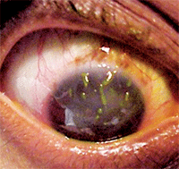
Her intraocular pressure measured 14mm Hg O.U. Dilated fundoscopy was within normal limits in both eyes. The pertinent anterior segment findings are illustrated in the photograph.
Your Diagnosis
How would you approach this case? Does this patient require any additional tests? What is your diagnosis? How would you manage this patient? What’s the likely prognosis?
Additional testing might have included a Schirmer screening to evaluate tear volume, tear quality analysis, sodium fluorescein, lissamine green and rose bengal dye tests to confirm and grade the corneal and conjunctival epitheliopathy, exophthalmometry to rule out proptosis (thyroid testing if positive) and eyelid strength testing and blink analysis to rule out lagophthalmos and cranial nerve VII palsy. Review of more extensive patient history could also help rule out the use of medications capable of causing dry eye, and laboratory testing could help rule out associated systemic etiologies.
The diagnosis in this case is dry eye and filamentary keratopathy secondary to poor eyelid closure, tear spread and tear quality in the patient’s right eye. The filaments were debrided and bandage contact lenses were prescribed along with copious lubrication therapy. The patient was scheduled for follow up in one week. At the first review visit, the condition had resolved completely and the patient reported no new symptoms or complications. The contact lens therapy was continue for one additional week along with the lubrication therapy and education was provided to augment tear production (flax seed oil, omega-3 supplements). At a follow up visit one week later, the patient was 100% resolved. The contact lens portion of the treatment was discontinued and the lubrication portion of the treatment was sustained. Oral vitamin C was also recommended secondary to its reputation to enhance collagen cross linkage.

An anterior segment view of our 57-year-old patient’s right eye. What is the underlying cause of this presentation?
Many collagen vascular diseases can result in unilateral or bilateral dry eye as well as noninfectious peripheral corneal inflammation. Rheumatoid arthritis (RA) and secondary Sjögren's syndrome are the most common systemic causes of dry eye disease. Other differential diagnoses of dry eye include poor lid-to-globe congruity, poor lid closure secondary to CN VII palsy, radiation exposure, chemical exposure, Sjögren's syndrome, severe atopic keratoconjunctivitis, scleroderma, erythema multiforme, trachoma, Beçhet’s disease and sarcoidosis.1-4
Medications, such as systemic birth control pills, practolol, topical epinephrine, idoxuridine, echothiophate iodide, pilocarpine, timolol and dipivefrin, have also been implicated. The combination of punctate epithelial keratitis and increased mucus production are the necessary ingredients for filamentary keratitis.5-7
Acetylcysteine is one of the most common treatments for tenacious mucus deposits that adhere to the superior eyelid cobblestones or the cornea, as thick, ropy strands. This medication is known to break the disulfide bonds and dissolve these deposits, and is effective for all three types of excessive mucus. Acetylcysteine is commercially available as Mucomyst (Bristol-Myers Squibb), which should be applied q.i.d.8-13
Other mainstay treatments include the use of ocular surface lubricants (e.g., artificial tears and/or ointments) or pressure patches to improve patient comfort and reduce the frictional effects of eyelid blinking. Also, hypertonic sodium chloride may have a beneficial effect. The clinician, depending upon disease severity and patient symptoms, can provide antimicrobial prophylaxis and cycloplegics. Finally, behavior modification may train patients to open their eyes slowly upon awakening.8-13
In cases that involve bigger erosions with loose sheets of epithelium, scraping and debridement with a Weck-cel sponge, Kimura spatula or saturated, cotton-tipped applicator may help induce efficient healing. Extended-wear bandage soft contact lenses may provide comfort and support the healing process, with minimal compromise of vision. Bandage lenses have the additional benefit of protecting and isolating the fragile, healing epithelium from the disruptive “windshield-wiper effect” of blinking. These lenses should remain on the cornea for at least six to eight weeks to allow the basement membrane and hemidesmosomes sufficient time to reorganize. A collagen shield may be a useful alternative for short-term relief and epithelial support. Additionally, oral acetaminophen and ibuprofen can provide adequate analgesia.9,14,15
If the erosions are severe or frequent, anterior stromal micropuncture or laser superficial keratectomy may be potential therapeutic options. Punctal occlusion with plugs or cautery can be performed to increase the lacrimal lake if scarring does not occur naturally. Any blepharitis may be controlled with regular lid scrubs and antibiotic drops or ointments. In worst-case scenarios, mucous membrane grafts can be used to replace irreparably scarred conjunctivae. Systemic immunosuppressive agents, such as cyclosporin, cyclophosphamide, Imuran (azathioprine, GlaxoSmithKline) and topical corticosteroids, can also be used to control the condition.8,16
1. Gregory DG. The ophthalmologic management of acute Stevens-Johnson syndrome. Ocul Surf. 2008;6(2):87-95.
2. Guzey M, Karaman SK, Satici A, et al. Efficacy of topical cyclosporine A in the treatment of severe trachomatous dry eye. Clin Experiment Ophthalmol. 2009;37(6):541-9.
3. Lemp MA. Evaluation and differential diagnosis of keratoconjunctivitis sicca. J Rheumatol Suppl. 2000;61(12):11-4.
4. Heiligenhaus A, Wefelmeyer E, Schrenk M. Tear-film deficiencies in patients with sarcoidosis; clinical study of 56 patients. Klin Monbl Augenheilkd. 2002;219(7):502-6.
5. Apostol S, Filip M, Dragne C, et al. Dry eye syndrome. Etiological and therapeutic aspects. Oftalmologia. 2003;59(4):28-31.
6. Stewart WC, Stewart JA, Nelson LA. Ocular surface disease in patients with ocular hypertension and glaucoma. Curr Eye Res. 2011;36(5):391-8.
7. Santaella RM, Fraunfelder FW. Ocular adverse effects associated with systemic medications: recognition and management. Drugs. 2007;67(1):75-93.
8. Diller R, Sant S. A case report and review of filamentary keratitis. Optometry 2005; 76(1):30-6. (same as 18)
9. Albietz, J, Sanfilippo P, Troutbeck R, et al. Management of filamentary keratitis associated with aqueous-deficient dry eye. Optom Vis Sci. 2003; 80(6):420-30. (same as 14)
10. Pons ME, Rosenberg SE. Filamentary keratitis occurring after strabismus surgery. J AAPOS. 2004;8(2):190-1.
11. Kakizaki H, Zako M, Mito H, et al. Filamentary keratitis improved by blepharoptosis surgery: two cases. Acta Ophthalmol Scand. 2003;81(6):669-71.
12. Tabery HM. Filamentary keratopathy: a non-contact photomicrographic in vivo study in the human cornea. Eur J Ophthalmol. 2003;13(7):599-605.
13. Cher I. Blink-related microtrauma: when the ocular surface harms itself. Clin Experiment Ophthalmol. 2003;31(3):183-90.
14. Arora I, Singhvi S. Impression debridement of corneal lesions. Ophthalmology. 1994;101(12):1935-40.
15. Hadassah J, Prakash D, Sehgal PK et al. Clinical evaluation of succinylated collagen bandage lenses for ophthalmic applications. Ophthalmic Res. 2008;40(5):257-66.
16. Perry HD, Doshi-Carnevale S, Donnenfeld ED, et al. Topical cyclosporine A 0.5% as a possible new treatment for superior limbic keratoconjunctivitis. Ophthalmology. 2003;110(8):1578-81.

