Photo Atlas of Ocular DiseaseReview of Optometry has created the following new resource—a photo atlas of ocular diseases across the entire spectrum of eye care. In addition to over 200 high-quality photos, you'll find clinical pearls for each condition and links to relevant articles that provide detailed guidance. An index of the entire atlas can be found here or you can check out the conditions featured in their respective categories: |
“Start on the outside and work your way in” has been the mantra of optometric educators, and that includes examination of the conjunctiva, cornea and iris. While it is easy to breeze by these structures on the way to the retina, the anterior segment holds vital information on the well-being of the patient. Inflammation, infection and malignancy may point to greater systemic concerns, as well as sight-threatening disease.
Under low magnification, the bulbar conjunctiva/sclera should appear white and moist. Non-wetting areas may point to exposure, vitamin deficiencies, functional eyelid anomalies or systemic autoimmune diseases. Color changes could reflect liver conditions (yellow), ultraviolet/exposure damage (pinguecula, yellow), the potential for malignancy (primary acquired melanosis, brown; squamous cell carcinoma, pink). Dilated vessels, as well as chemosis, indicate inflammation and the root cause—such as infection, immune or dry eye—should be sought out.
The papillary conjunctiva should be free of inflammation and structural changes as well.
The visual examination of the cornea should go from the epithelium to the endothelium, as well as scanning the limbus 360 degrees. The cornea should be free of vessels (deep and superficial), infiltrates and foreign body material/deposits. It should have a uniform thickness, without thinning, edema, striae, folds or guttae. Additional testing with topography, OCT and specular microscopy are helpful for closer examination and to meet diagnostic criteria for many anterior segment diseases.
Like the cornea, the iris also benefits from both visual and tomographic examinations—for instance, to rule out neovascular and malignant lesions. In addition, gonioscopy is helpful for viewing the angle for the eye, which is a necessity for glaucoma management, post trauma evaluation and tumor concerns.
The anterior segment is easy to photodocument for comparison and communication. Below, we share dozens of images of the disorders that can befall the structures of the anterior segment from a vast array of etiologies.
Feel free to click the condition that interests you to jump to it, or scroll to look through all the conditions.
Epithelial basement membrane dystrophy
Central corneal dystrophy of François
Fuchs’ endothelial corneal dystrophy
Terrien’s marginal degeneration
Peripheral ulcerative keratitis
Conjunctival intraepithelial neoplasm
Giant papillary conjunctivitis
Ocular surface squamous neoplasia
Limbal stem cell deficiency
 |
| Photo: Alison Bozung, OD. Click image to enlarge. |
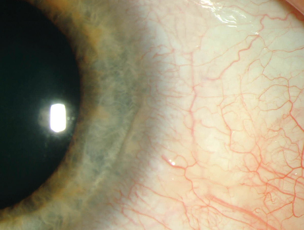
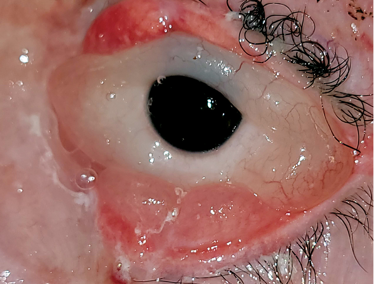
LSCD occurs when damage to the limbal niche allows for conjunctivalization of the cornea.
Suggested reading:
Corneoscleral Concerns: Trouble at the Border
Transplantation for Limbal Stem Cell Deficiency
Neurotrophic keratitis
 |
| Photo: Alison Bozung, OD. Click image to enlarge. |
Characterized by poor epithelial healing and reduced sensation, neurotrophic keratitis results from damage to corneal nerves.
Suggested reading:
Take On Neurotrophic Keratitis With These Clinical Tools
Arcus senilis
 |
| Photo: Paul Karpecki, OD. Click image to enlarge. |
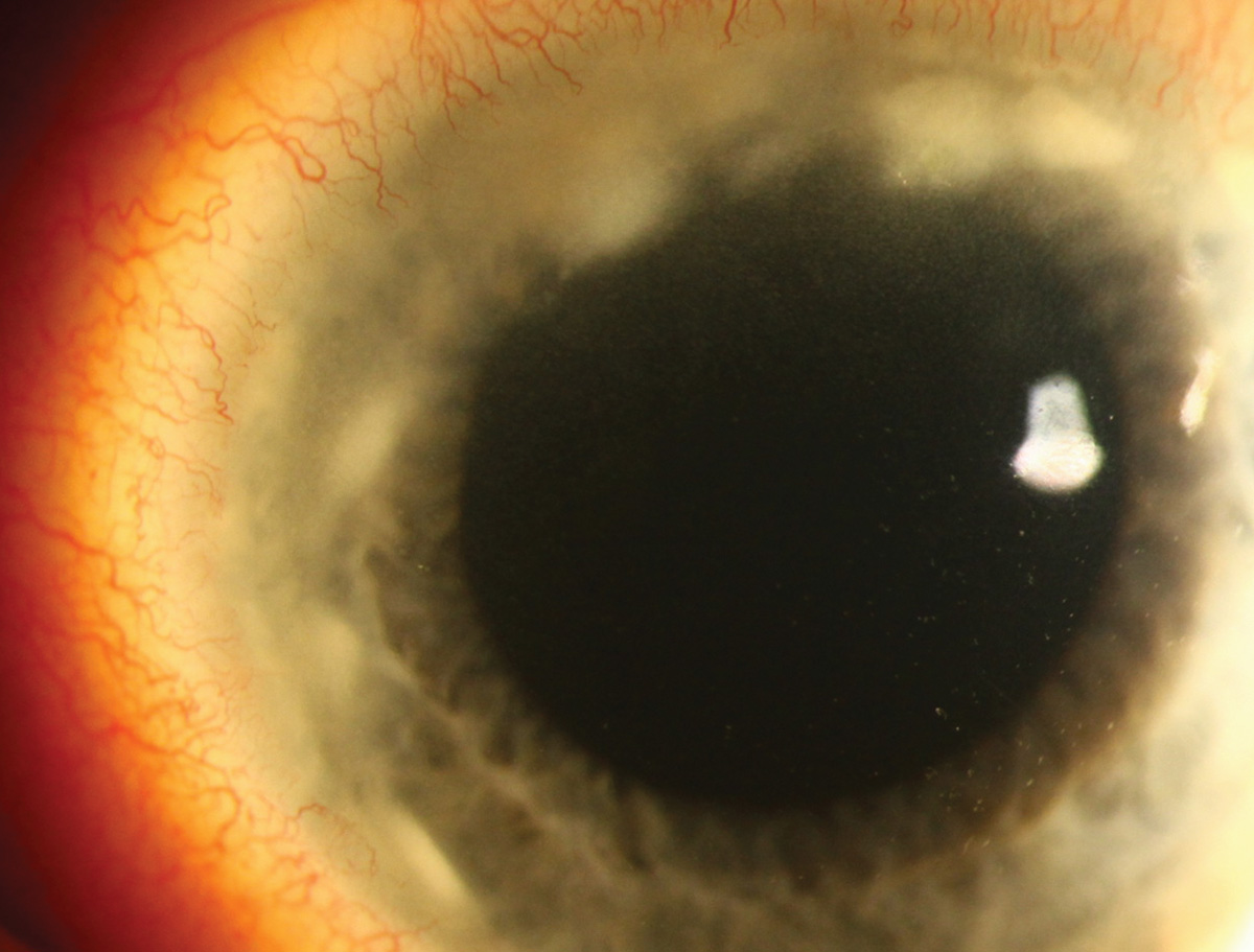
In arcus senilis, age-related deposition of lipids occurs in response to increased permeability of limbal blood vessels.
Suggested reading:
Epithelial basement membrane dystrophy
 |
| Photos: Mitch Ibach, OD. Click image to enlarge. |
 |
| Click image to enlarge. |
Epithelial basement membrane dystrophy, considered more of a degenerative condition due to its weak genetic association, presents with grayish dots and lines (first image). Thickened epithelium may be noted (second image, from a different patient).
Suggested reading:
Unlocking the Mystery of Corneal Dystrophies
Lisch dystrophy
 |
| Photo: Aaron Bronner, OD. Click image to enlarge. |
Lisch dystrophy produces feathery clusters of microcystic epithelial tissue in a whorled pattern sweeping centrally from the limbus.
Suggested reading:
Unlocking the Mystery of Corneal Dystrophies
Schnyder’s dystrophy
 |
| Photo: Suzanne Sherman, OD. Click image to enlarge. |
Schnyder’s dystrophy presents in the first decade of life with central disciform opacities often surrounded by a dense arcus.
Suggested reading:
Corneal Dystrophies Front to Back
Lattice dystrophy
 |
| Photo: Paul Karpecki, OD. Click image to enlarge. |
 |
| Photo: University of Iowa. Click image to enlarge. |
Lattice dystrophy represents deposition of amyloid in the anterior stroma, which forms a characteristic branching linear pattern. Most cases are inherited as an autosomal dominant trait (type I), but a rarer form arises from systemic amyloidosis (type II).
Suggested reading:
Granular dystrophy
 |
| Photo: Stephanie Fromstein, OD. Click image to enlarge. |
 |
| Photo: Mitch Ibach, OD. Click image to enlarge. |
In granular dystrophy type I (first photo), hyaline deposits form in the anterior stroma, eventually becoming more confluent as it progresses. Type II or Avellino dystrophy (second photo) also features lattice-like lesions of the mid-posterior stroma; however, these branches do not cross.
Suggested reading:
Corneal Dystrophies Front to Back
Reis-Bucklers
 |
| Photo: University of Iowa. Click image to enlarge. |
Reis-Bucklers causes reticular opacities in Bowman’s that become less discrete over time, later extending into the stroma.
Suggested reading:
Unlocking the Mystery of Corneal Dystrophies
Central corneal dystrophy of François
 |
| Photo: Suzanne Sherman, OD. Click image to enlarge. |
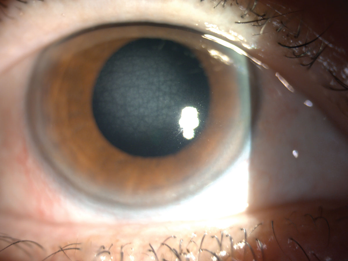
CCDF possesses autosomal dominant inheritance with onset during the first or second decade of life. Often, it is described as appearing similar to crocodile shagreen. Signs include translucent, scaly polygonal or rounded opacities residing deep within the stroma and are surrounded by clear tissue. It has also been described as a fluffy opacity, appearing as cracked ice.
Suggested reading:
Fuchs’ endothelial corneal dystrophy
 |
| Photos: University of Iowa. Click image to enlarge. |
 |
| Click image to enlarge. |
 |
| Click image to enlarge. |
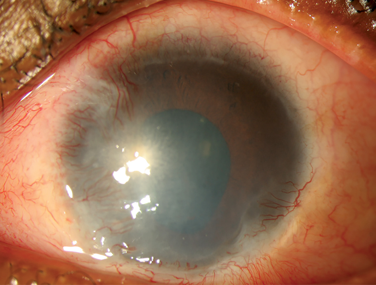
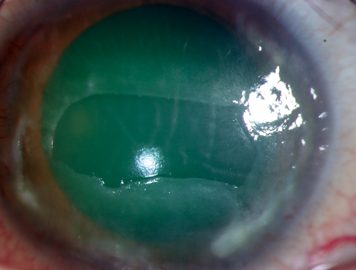
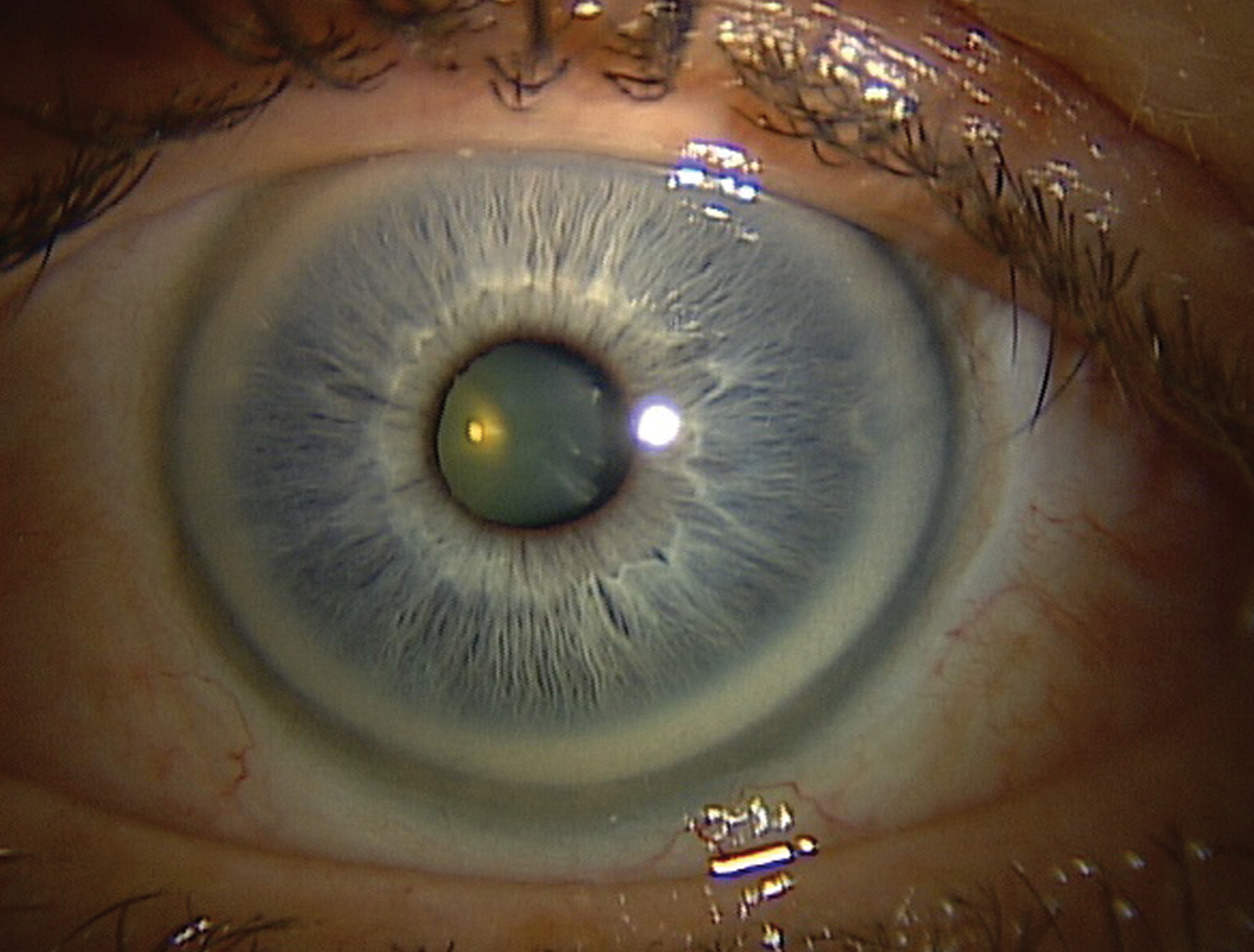
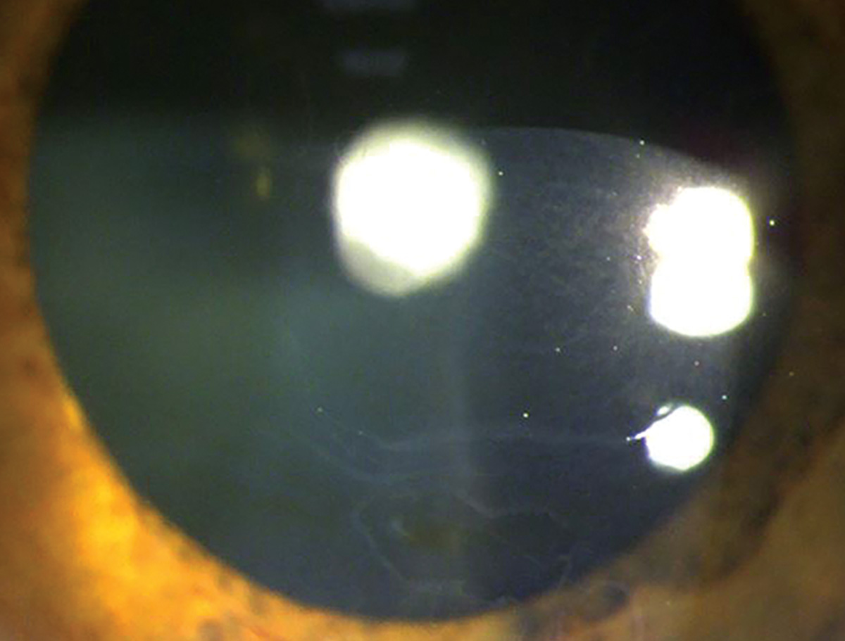
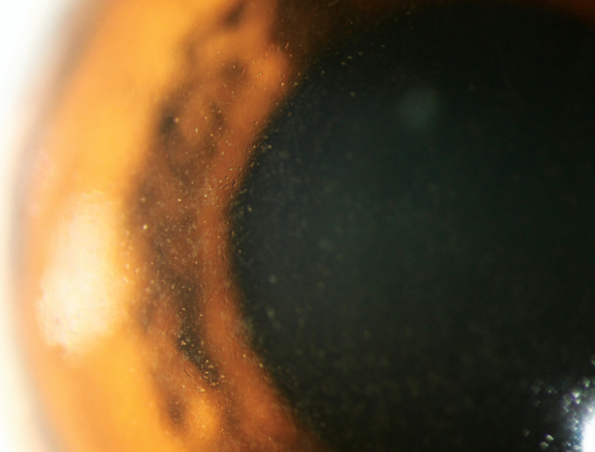
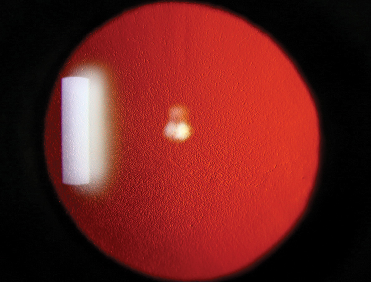
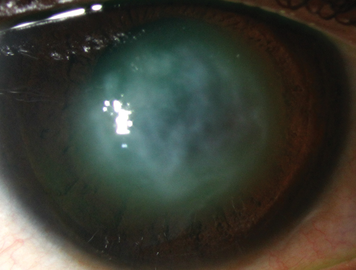
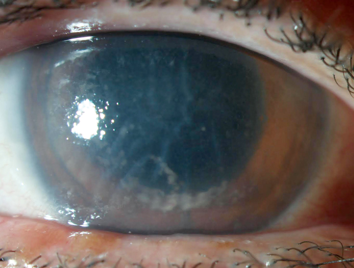
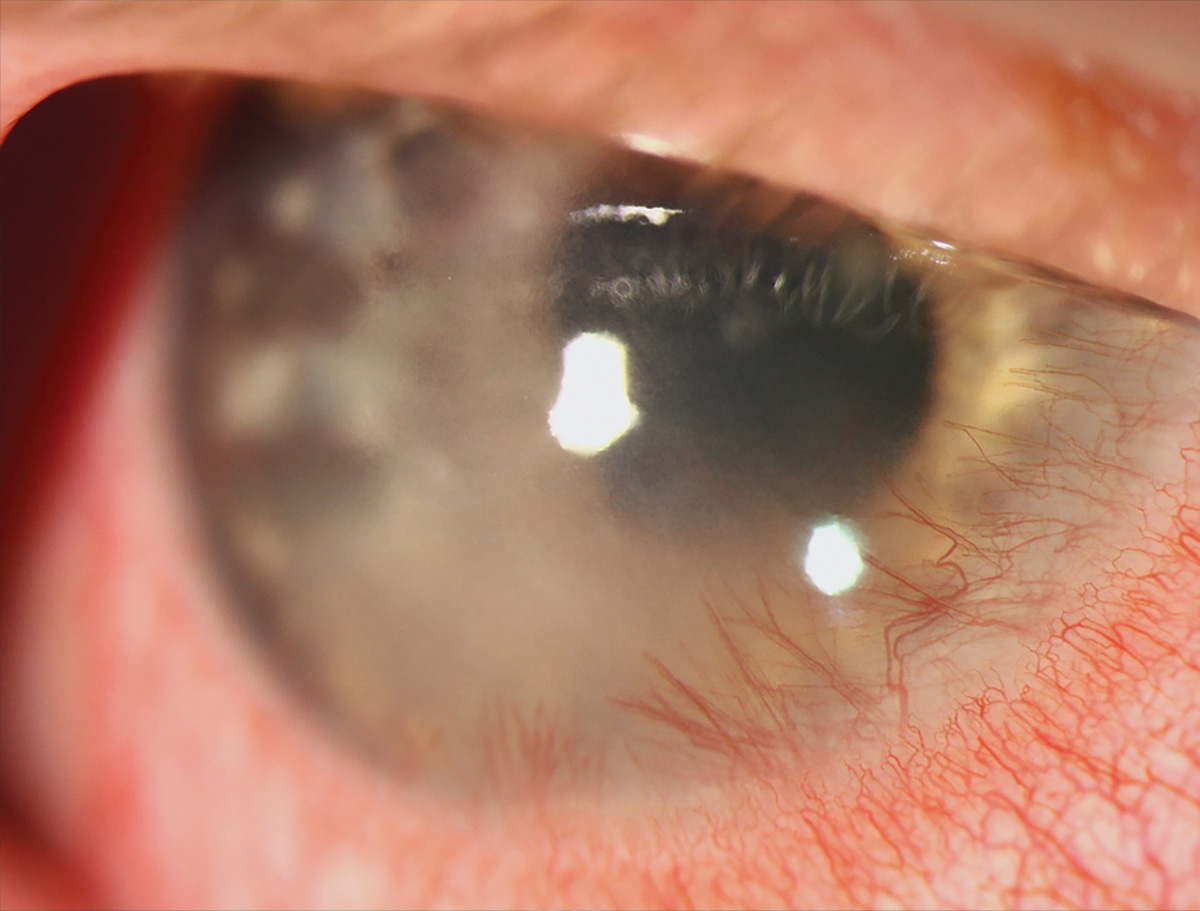
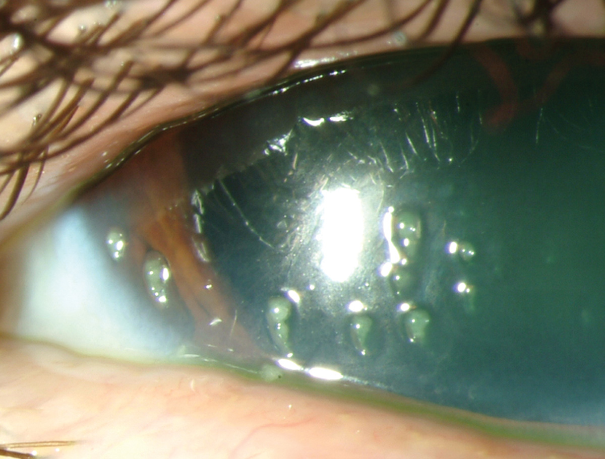
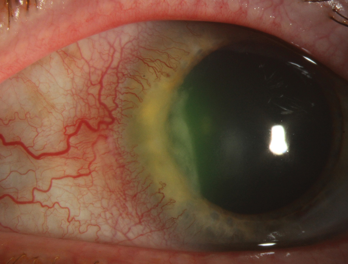
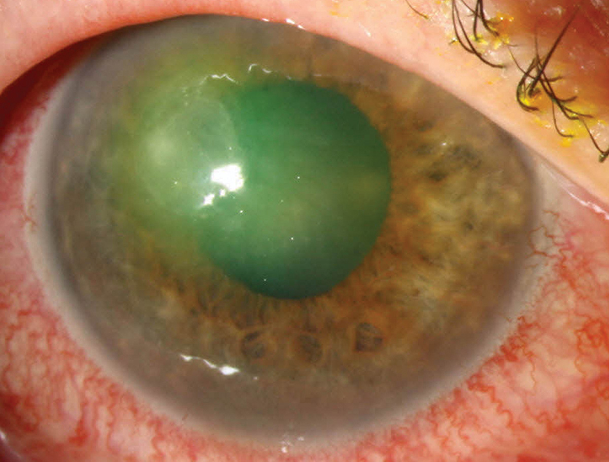
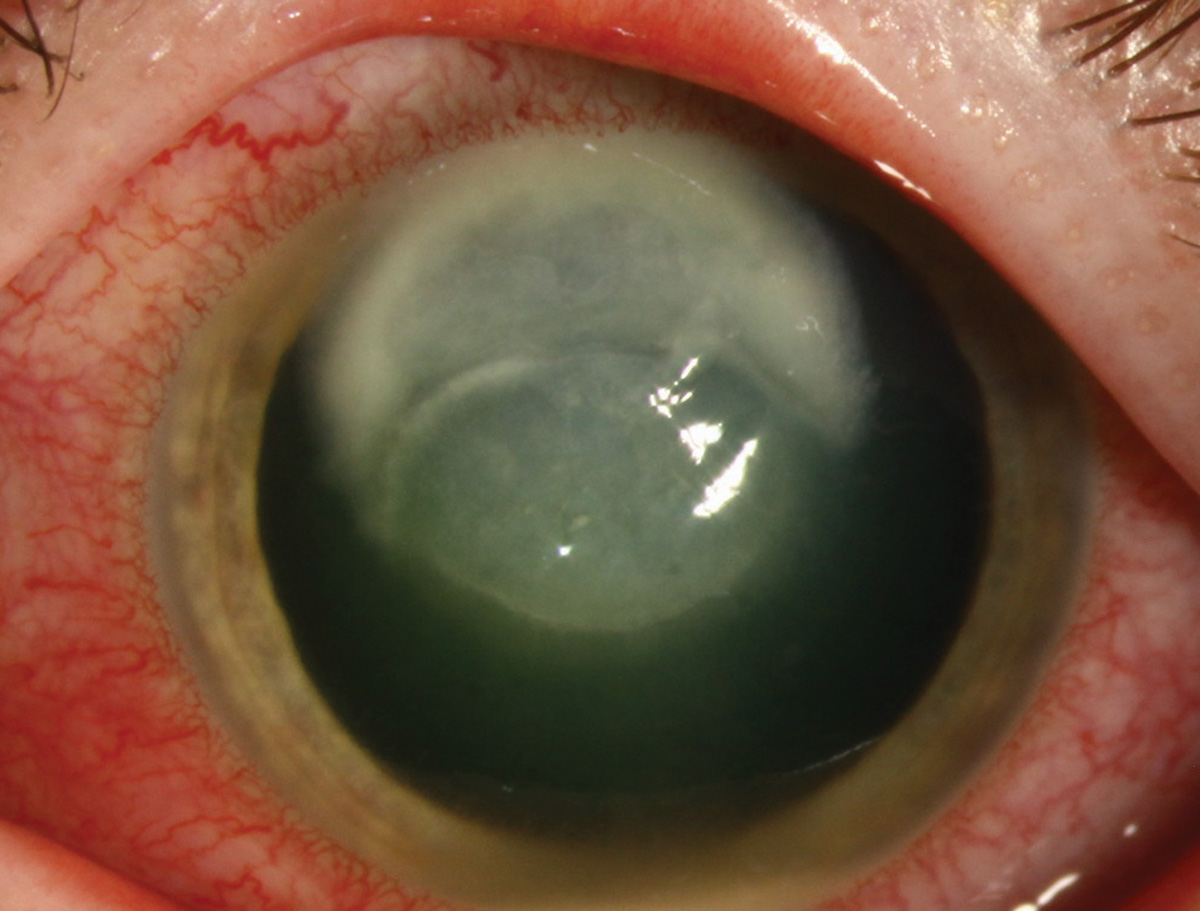
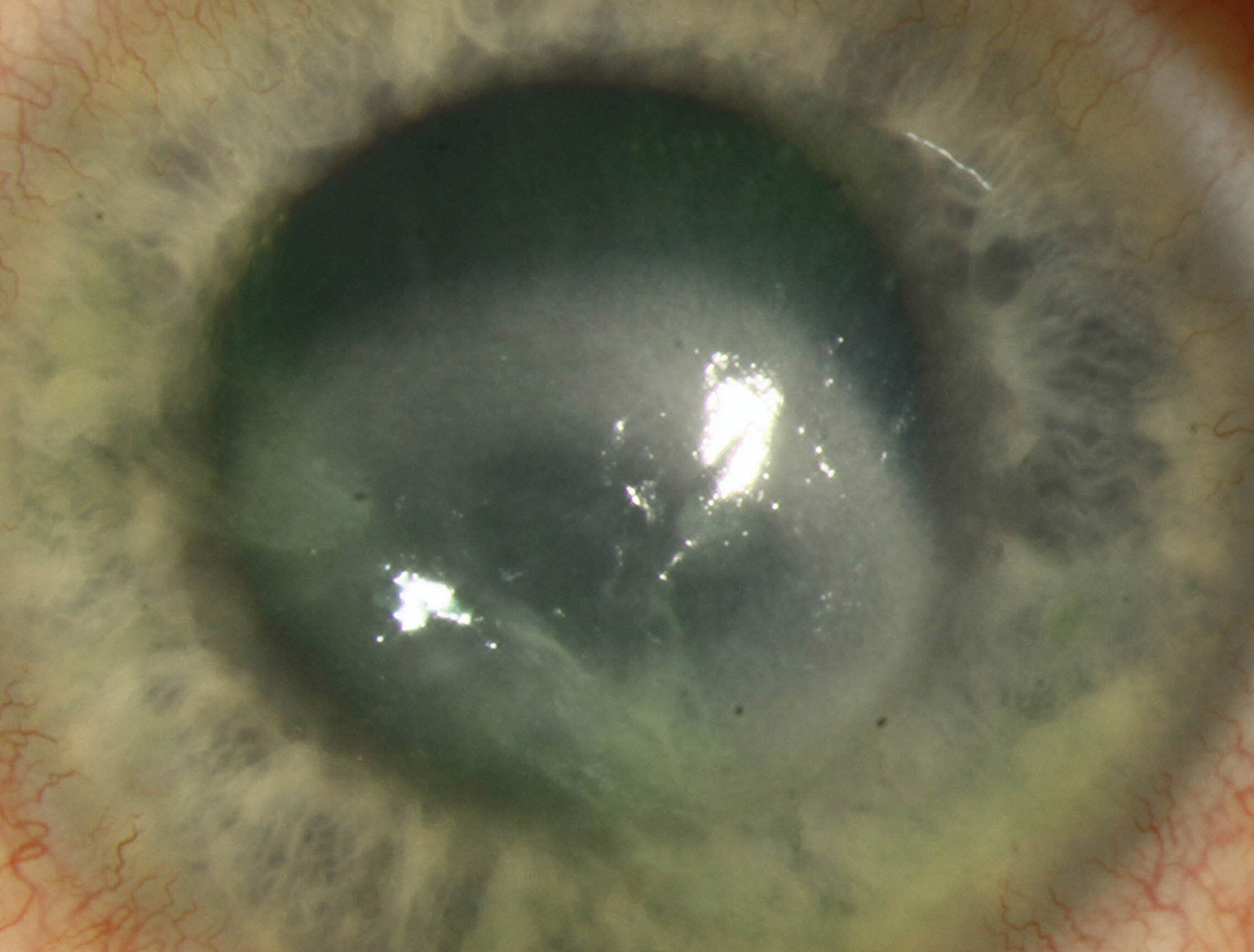
Fuchs’ endothelial corneal dystrophy (FECD), a bilateral genetic dystrophy characterized by progressing guttae leading to corneal edema and blurred vision. Patients may complain of worse vision in the morning, halos, dry eye and photophobia. In late-stage FECD with chronic corneal edema, patients may develop painful bullae.
Suggested reading:
Terrien’s marginal degeneration
 |
| Photos: Rami Aboumourad, OD. Click image to enlarge. |
 |
| Click image to enlarge. |
 |
| Click image to enlarge. |
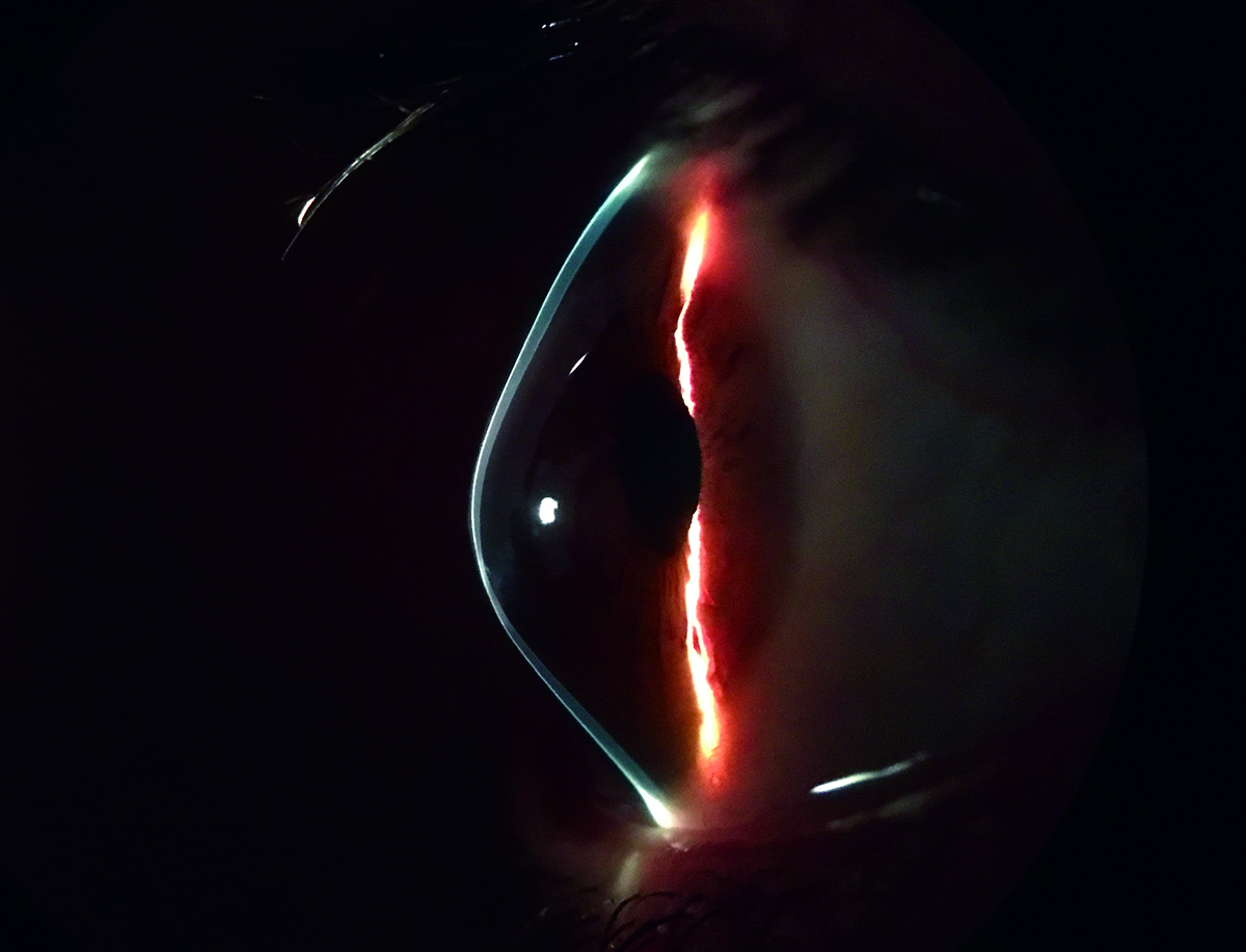
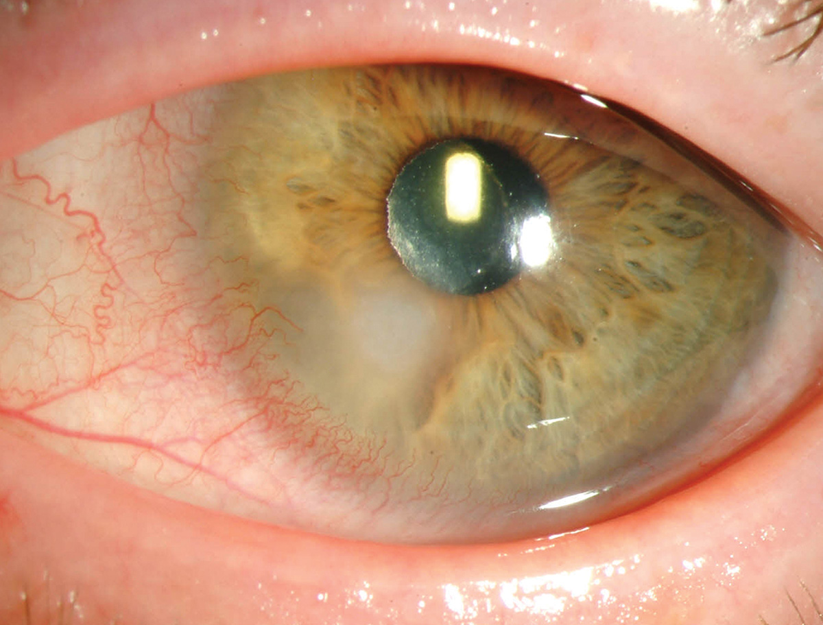
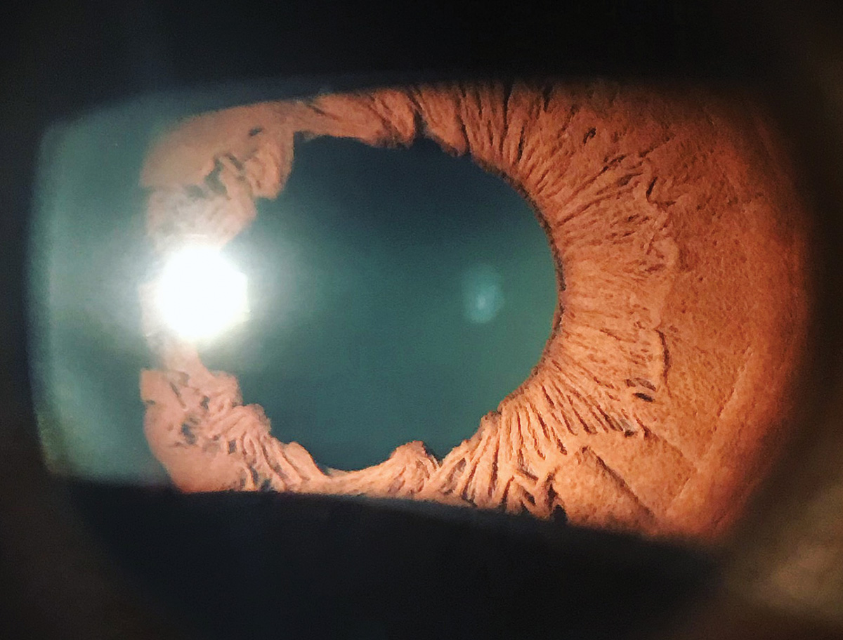
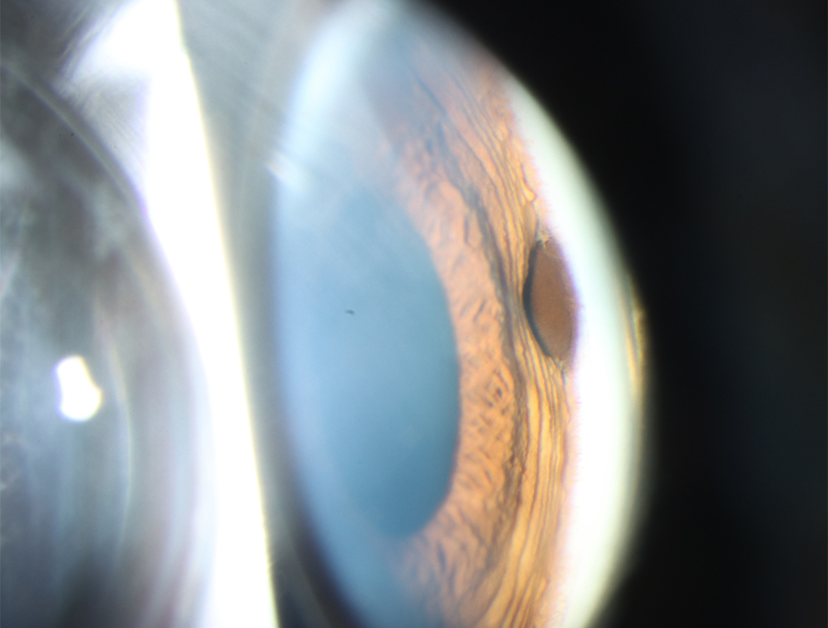
Terrien’s marginal degeneration is a progressive peripheral corneal disorder characterized by a paucity of inflammation in the setting of painless corneal thinning. Most patients have superior thinning with stromal lipid deposition and an overlying intact epithelium. Patients often develop high corneal astigmatism and may develop complications such as corneal perforation.
Suggested reading:
Corneoscleral Concerns: Trouble at the Border
Keratoconus
 |
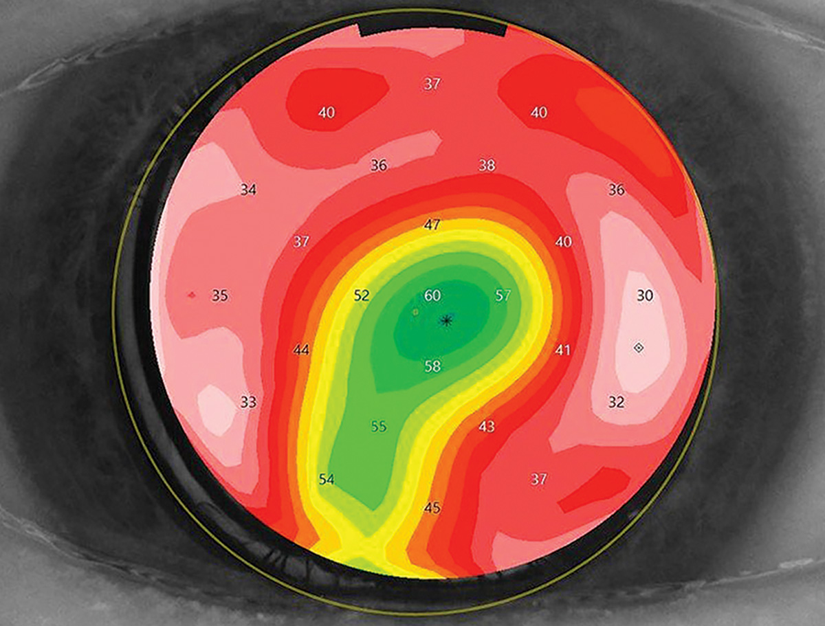
| Photo: Irving Martinez-Navé, OD. Click image to enlarge. |
 |
| Click image to enlarge. |
In keratoconus, thinning of the cornea produces anterior bulging in a conical form, sometimes visible on gross examination (first photo). At the slit lamp, look for Munson’s sign (second photo), as well as Vogt’s striae and Fleischer rings.
Suggested reading:
A New Consensus on Keratoconus
Trends, Challenges and Controversies in Keratoconus
Keratoconus: What Surprises the Experts?
Hydrops
 |
| Photo: Alison Bozung, OD. Click image to enlarge. |
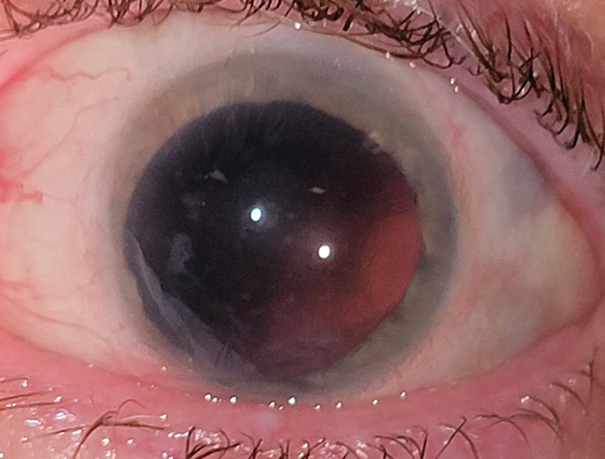
Acute corneal hydrops occurs when a break in Descemet’s membrane allows for stromal edema, seen here in keratoconus.
Suggested reading:
Mooren’s ulcer
 |
| Photo: University of Iowa. Click image to enlarge. |
Mooren’s ulcer, a circumferential corneal thinning near the limbus caused by immune-mediated eye disease.
Suggested reading:
Corneoscleral Concerns: Trouble at the Border
Lesson: Pathologic Causes of Irregular Astigmatism
Exposure keratopathy
 |
| Photo: Alison Bozung, OD. Click image to enlarge. |
Exposure keratopathy in this case resulted from a severe surgically induced cicatricial ectropion.
Suggested reading:
The Compromised Cornea: Take Cover
Dellen
 |
| Photos: University of Iowa. Click image to enlarge. |
 |
| Click image to enlarge. |
Dellen is a form of localized corneal thinning caused by desiccation. The condition is most commonly associated with contact lens wear or conjunctival masses, which disrupt the normal tear film.
Suggested reading:
Corneoscleral Concerns: Trouble at the Border
Graft-vs-host disease
 |
| Photo: University of Iowa. Click image to enlarge. |
Graft-vs-host disease in an allogenic bone marrow stem cell recipient with immune-mediated destruction of the ocular surface.
Suggested reading:
Graft-vs.-host Disease: How, Why and What Next
Verticillata
 |
| Photo: University of Iowa. Click image to enlarge. |
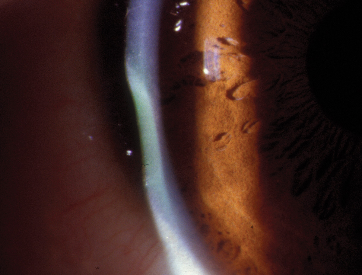
Verticillata from amiodarone use. Deposits form in the inferior corneal basal epithelium and branch out from a central whorl.
Suggested reading:
Don’t Be Scared by the Whirlwind
Know Your Systemic Meds: The Top 10 to Track
HSV keratouveitis
 |
|
Photo: Aaron Bronner, OD. Click image to enlarge. |
HSV keratouveitis is an uncommon but severe manifestation of corneal herpetic disease.
Suggested reading:
Herpetic Keratouveitis Front to Back
Be a Hero to Your HSVK Patients
HSV keratitis
 |
|
Photos: Alison Bozung, OD. Click image to enlarge. |
 |
| Click image to enlarge. |
Epithelial (first photo) and interstitial (second photo) HSV keratitis. Herpes simplex can take on many forms in the cornea including (but not limited to) classic “dendrites” as seen on the left image, stromal vascularization seen on the right and endotheliitis with corneal edema.
Suggested reading:
A Game Plan for Treating Corneal Infections
Be a Hero to Your HSVK Patients
Herpes Simplex Keratitis: Managing the Masquerader
Filamentary keratitis
 |
|
Photo: University of Iowa. Click image to enlarge. |
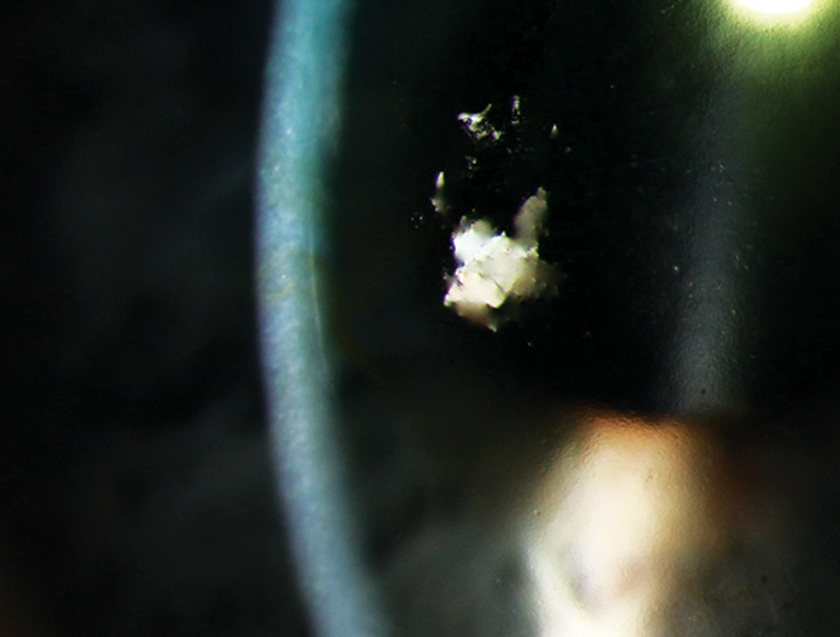
In filamentary keratitis, strands composed of epithelium, mucus and cellular debris form secondary to desiccation.
Suggested reading:
Fighting Filamentary Keratitis
Peripheral ulcerative keratitis
 |
| Photo: University of Iowa. Click image to enlarge. |
Peripheral ulcerative keratitis is an inflammation of the peripheral cornea with destruction of the epithelium and stroma.
Suggested reading:
Corneoscleral Concerns: Trouble at the Border
Understanding Corneal Ulcers and Infiltrates
Pseudomonas keratitis
 |
| Photos: University of Iowa. Click image to enlarge. |
 |
| Click image to enlarge. |
Pseudomonas keratitis (first photo) with hypopyon (second photo). This gram-negative pathogen often associated with contact lens wear presents with a ring of polymorphonuclear neutrophils around a central lesion.
Suggested reading:
A Game Plan for Treating Corneal Infections
Understanding Corneal Ulcers and Infiltrates
Lesson: Trends in Infectious Keratitis
Microbial keratitis
 |
| Photos: University of Iowa. Click image to enlarge. |
 |
| Click image to enlarge. |
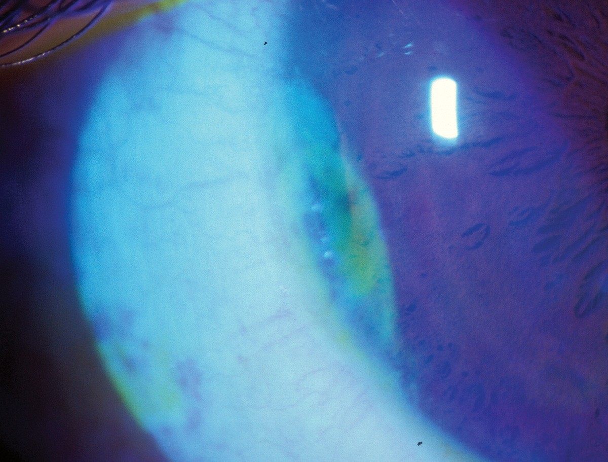
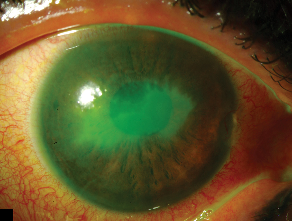
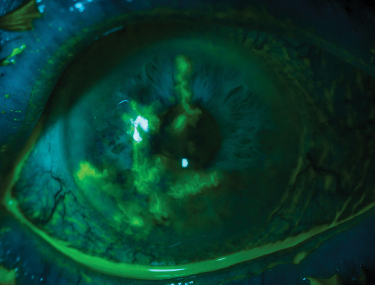
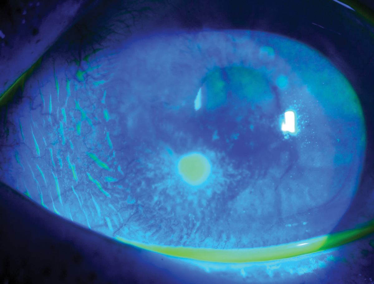
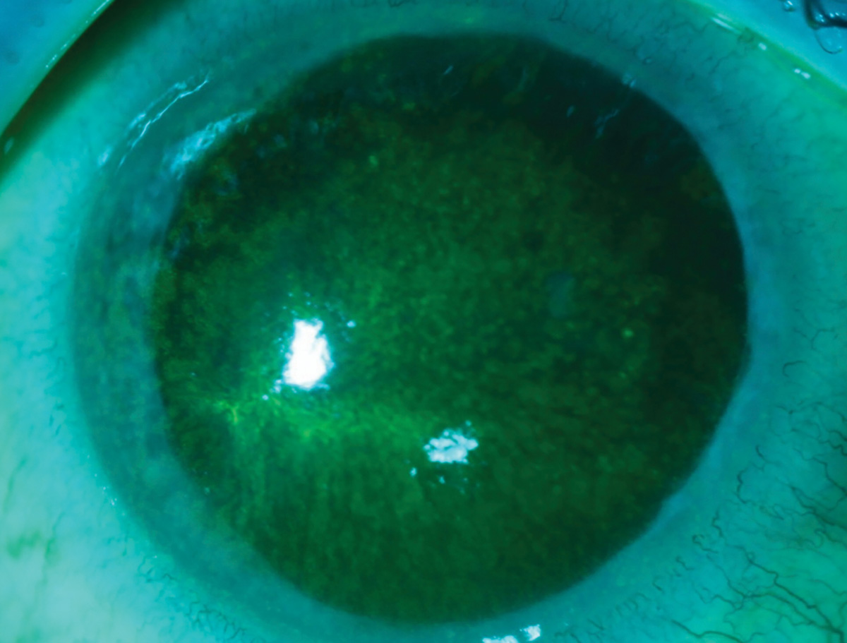
Microbial keratitis viewed with white light and fluorescein. In such cases, look for signs of a serious pathogen, including an infiltrate >1mm, presence of two or more lesions, central location, anterior chamber reaction and feathery borders.
Suggested reading:
A Game Plan for Treating Corneal Infections
The Dangers and the Diagnosis of CLMK
Marginal keratitis
 |
| Photo: Alison Bozung, OD. Click image to enlarge. |
This case of marginal keratitis has peripheral infiltrates with largely intact epithelium in a patient with blepharitis.
Suggested reading:
Understanding Corneal Ulcers and Infiltrates
The Dangers and the Diagnosis of CLMK
Acanthamoeba keratitis
 |
| Photos: University of Iowa. Click image to enlarge. |
 |
| Click image to enlarge. |
Suggested reading:
A Game Plan for Treating Corneal Infections
https://www.reviewofoptometry.com/article/contact-contaminators
The Dangers and the Diagnosis of CLMK
Fusarium ulcer
 |
| Photo: University of Iowa. Click image to enlarge. |
Consider the potential for fungal keratitis whenever multiple lesions are present.
Suggested reading:
A Game Plan for Treating Corneal Infections
Find Infectious Keratitis’s Root
Infiltrative Keratitis: Fight the Battle for Corneal Clarity
Pinguecula
 |
| Photo: University of Iowa. Click image to enlarge. |
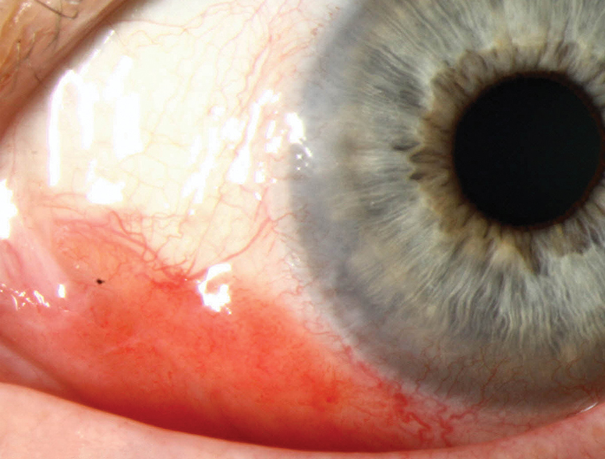
Pinguecula results from normal collagen replacement by thicker fibers when exposed to dryness and UV light.
Suggested reading:
Put a Red Eye Back in the Pink
Pterygium
 |
| Photo: University of Iowa. Click image to enlarge. |
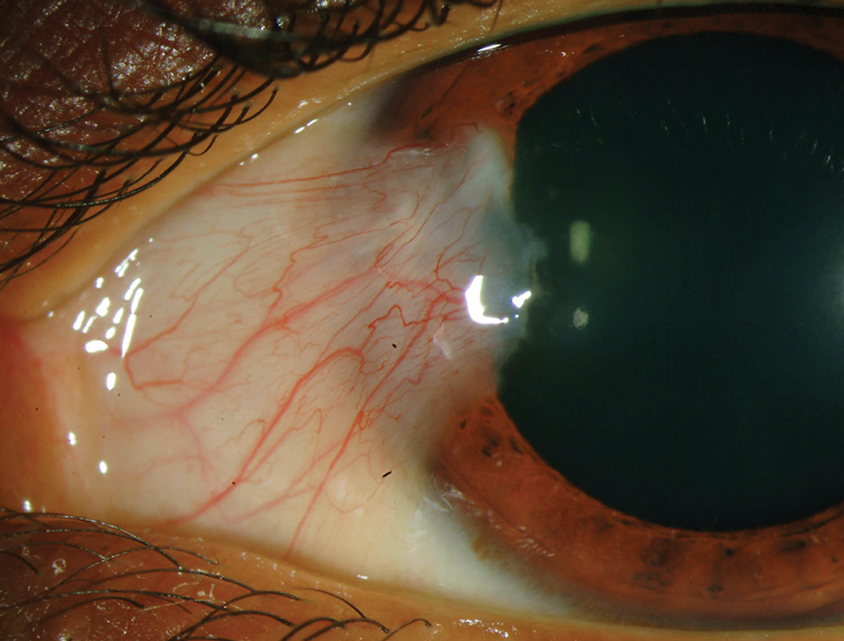
Pterygium, also a response to UV light exposure, shows fibrovascular tissue crossing the limbal border onto the cornea.
Suggested reading:
Addressing Pterygium in Optometric Practice
Corneal Optics are Significantly Affected by Pterygium
Limbal dermoid
 |
| Photo: University of Iowa. Click image to enlarge. |
Limbal dermoids are benign tumors that histologically may contain connective tissue, skin, fat, hair follicles and sweat glands.
Suggested reading:
Conjunctival intraepithelial neoplasm
 |
| Photos: Aaron Bronner, OD. Click image to enlarge. |
 |
| Click image to enlarge. |
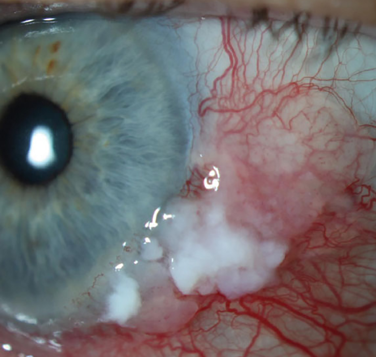
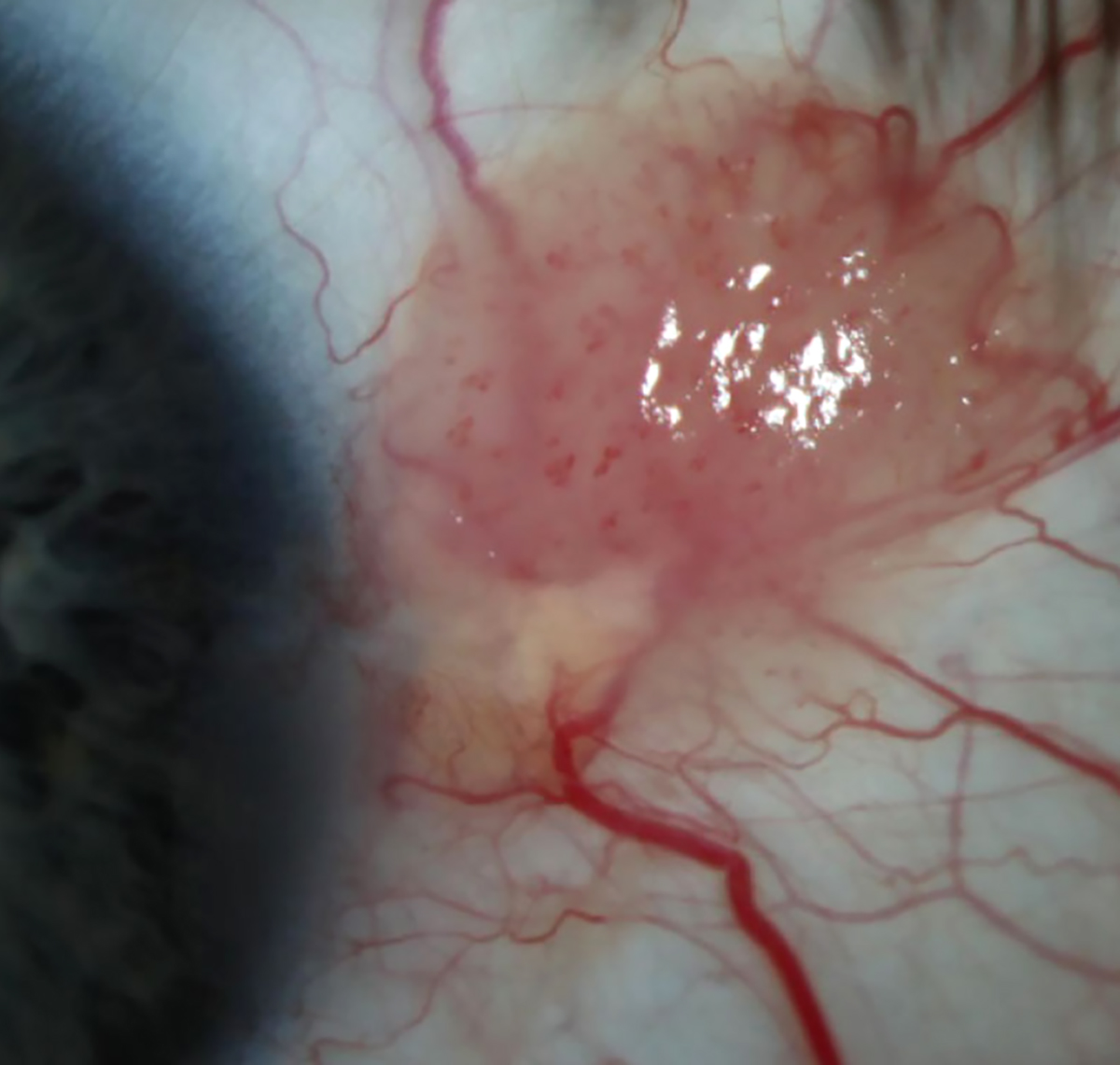
Conjunctival intraepithelial neoplasm (CIN), the most frequently encountered conjunctival neoplastic growth, is often misdiagnosed as more typical ocular surface growths like pinguecula or pterygium. These lesions are slowly progressive, locally invasive and have no metastatic potential. CIN is part of the spectrum of neoplastic disorders of the conjunctiva and cornea known collectively as ocular surface squamous neoplasia. If CIN becomes invasive by breaking through basement membrane, it is reclassified as invasive squamous cell carcinoma.
Suggested reading:
Is That Conjunctival Lesion Cancerous?
A Guide to Conjunctival Tumors
Pyogenic granuloma
 |
| Photo: University of Iowa. Click image to enlarge. |
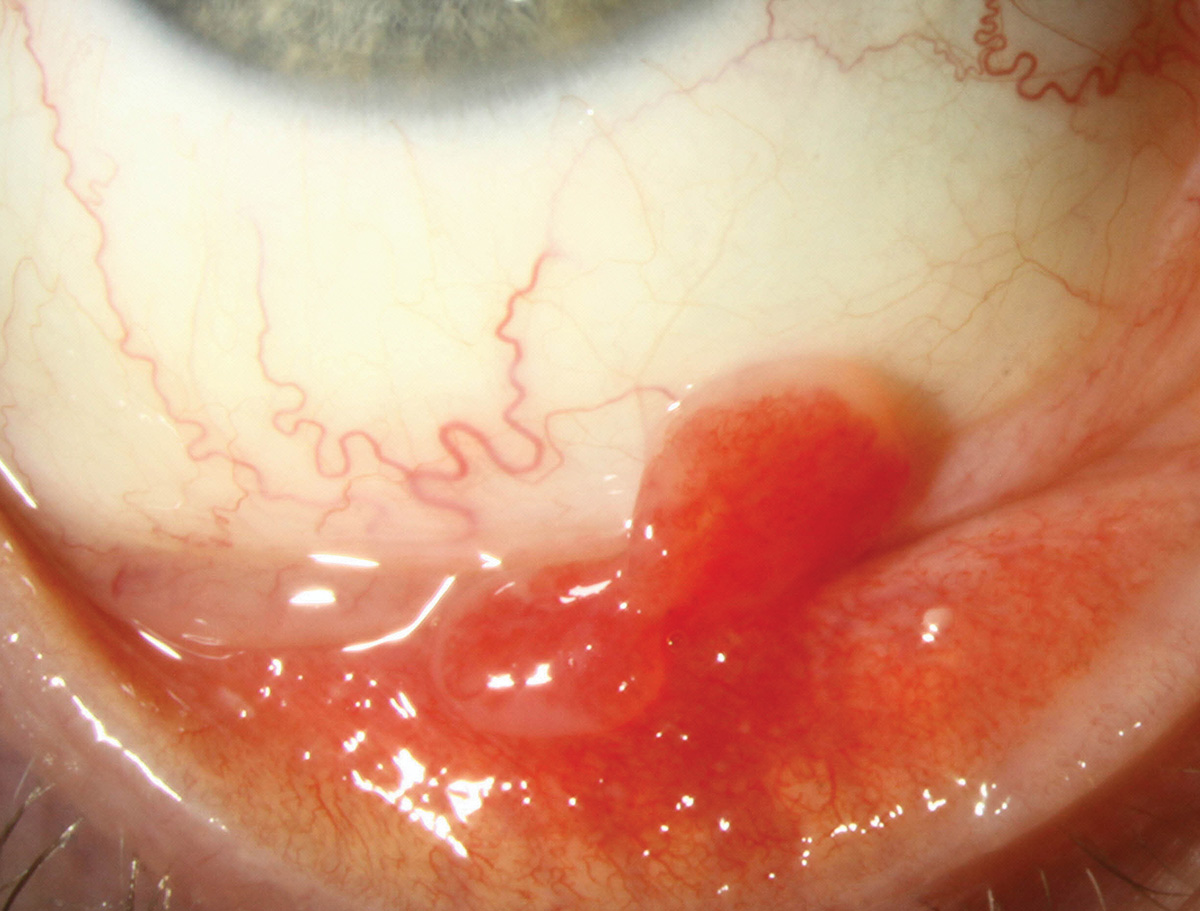
Pyogenic granuloma is a rapidly growing tissue hypertrophy secondary to an inflammatory agent.
Suggested reading:
Follicular conjunctivitis
 |
| Photo: University of Iowa. Click image to enlarge. |
Follicular conjunctivitis is an immunogenic activation of lymphoid follicles associated with many pathogens, drugs and allergies.
Suggested reading:
The Conjunctiva in Crisis: Ocular Irritation Unmasked
Conjunctivitis: Making the Call
Conjunctival cyst
 |
| Photo: University of Iowa. Click image to enlarge. |
Conjunctival cysts are harmless, painless fluid-filled vacuoles found on the ocular conjunctiva.
Suggested reading:
Giant papillary conjunctivitis
 |
| Photo: University of Iowa. Click image to enlarge. |
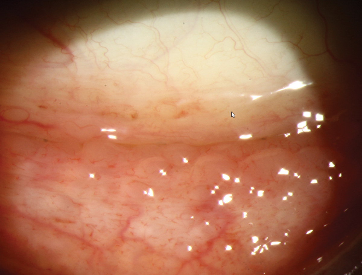
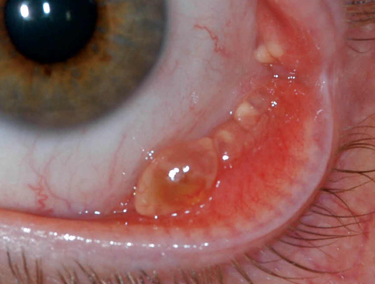
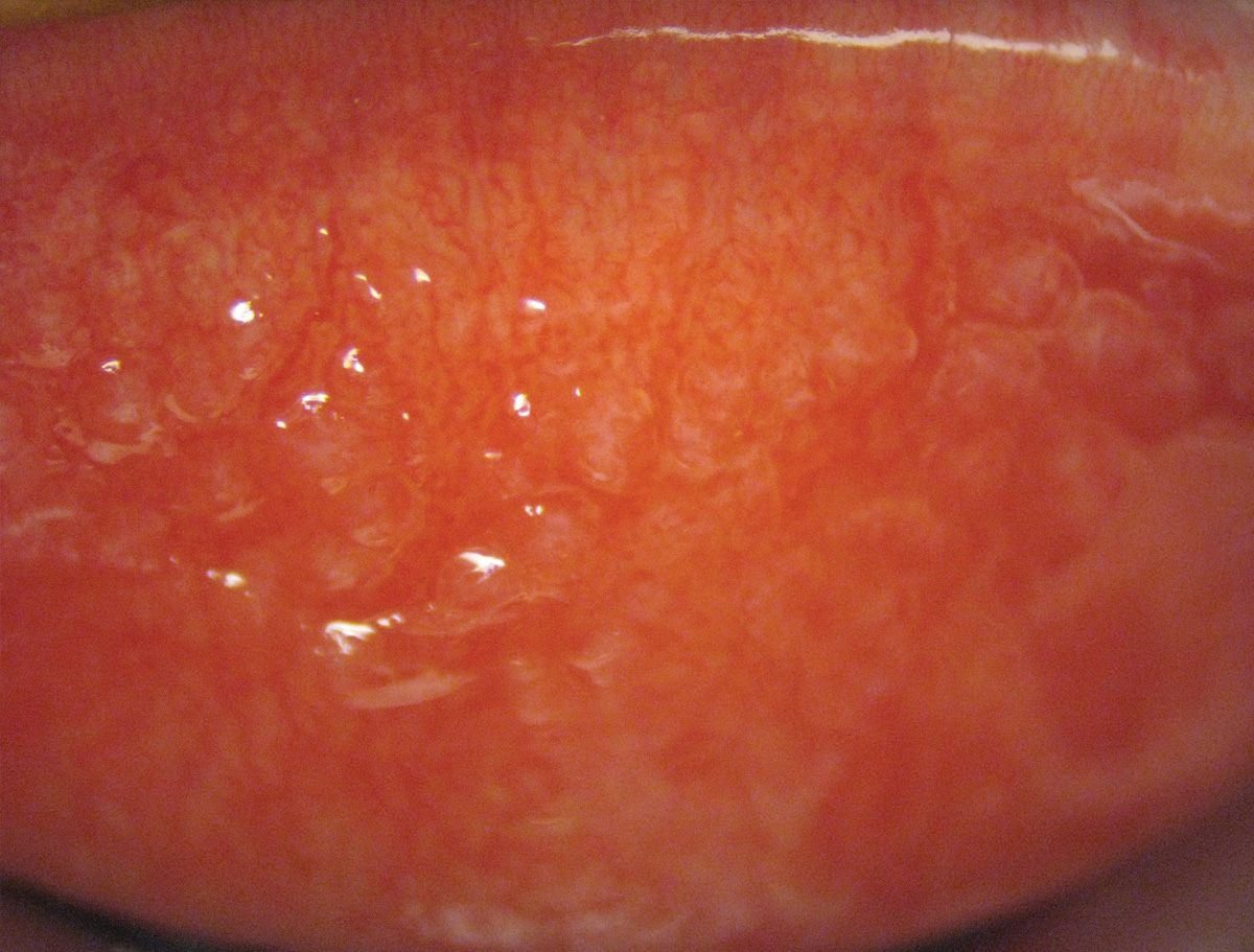
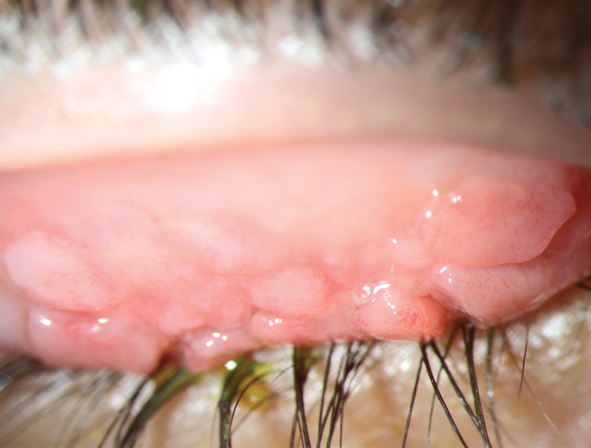
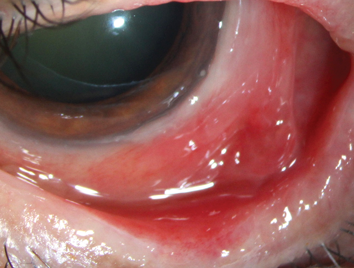
GPC, characterized by papillary hypertrophy of the superior tarsal conjunctiva, is on the decline thanks to daily disposable contact lens use, but can manifest in allergy cases.
Suggested reading:
Rethinking GPC: A New Look at an Old Problem
Vernal keratoconjunctivitis
 |
| Photos: Alison Bozung, OD. Click image to enlarge. |
 |
| Click image to enlarge. |
Cobblestone-like papillae (first photo) are a hallmark of vernal keratoconjunctivitis. This teenage patient endorsed intense itching and ocular discomfort. The patient also had developed a small shield ulcer and had small Horner-Trantas dots visible at the limbus (second photo).
Suggested reading:
Conjunctivitis: Making the Call
Subconjunctival hemorrhage
 |
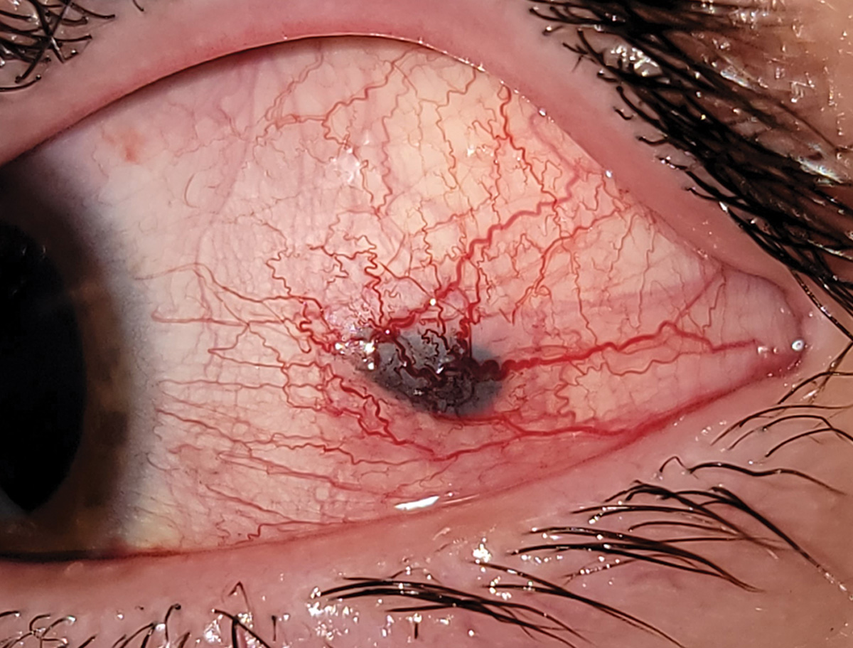
| Photo: University of Iowa. Click image to enlarge. |
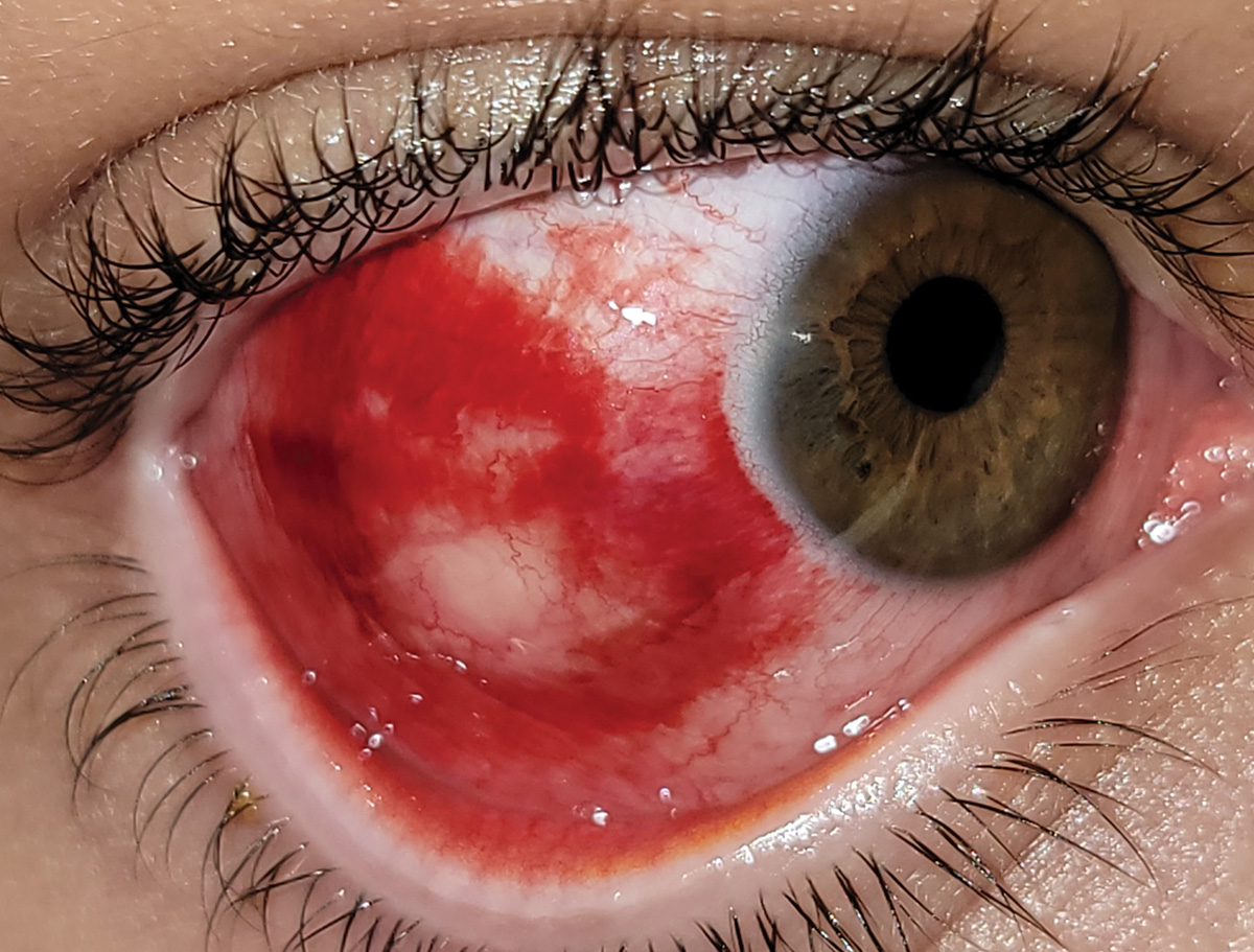
Subconjunctival hemorrhage is a benign finding of trapped blood commonly from Valsalva maneuvers or anticoagulant use.
Suggested reading:
Red Eye Remedies: New and Tried-and-True
Scleral thinning
 |
| Photo: University of Iowa. Click image to enlarge. |
Scleral thinning in HSV scleritis. Differential includes congenital/degenerative diseases, immunocompromise, infection and trauma.
Suggested reading:
My Patient Has Scleritis...Now What?
Gonorrhea conjunctivitis
 |
| Photo: Alison Bozung, OD. Click image to enlarge. |
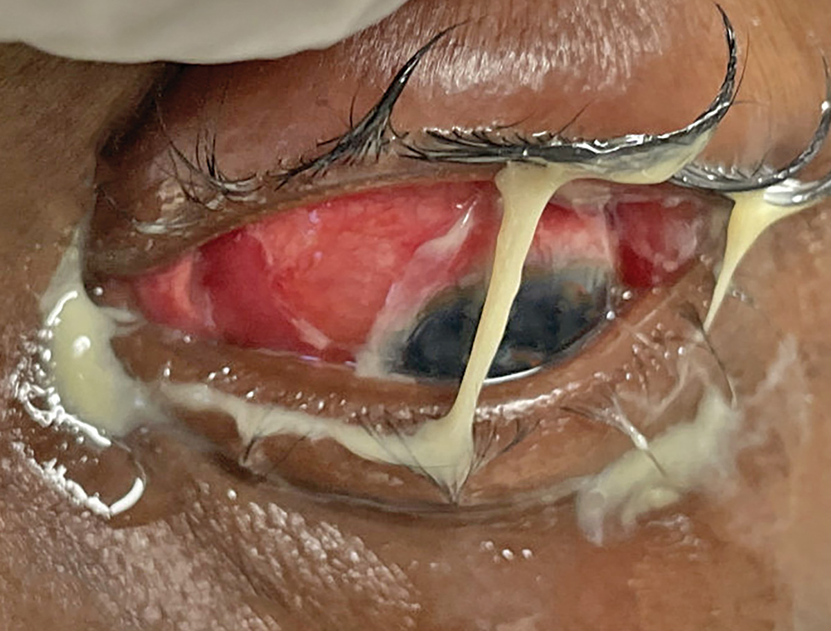
Gonorrhea conjunctivitis is a hyper-purulent conjunctivitis with rapid onset. Patients require systemic treatment.
Suggested reading:
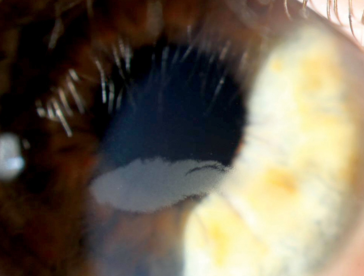
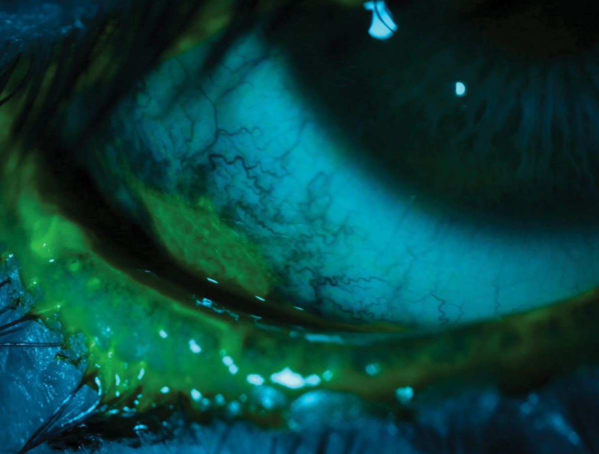
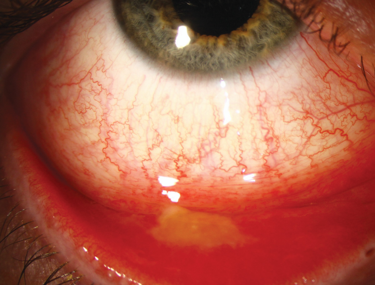
Ocular surface squamous neoplasia
 |
| Photos: Alison Bozung, OD. Click image to enlarge. |
 |
| Click image to enlarge. |
 |
| Click image to enlarge. |
 |
| Click image to enlarge. |
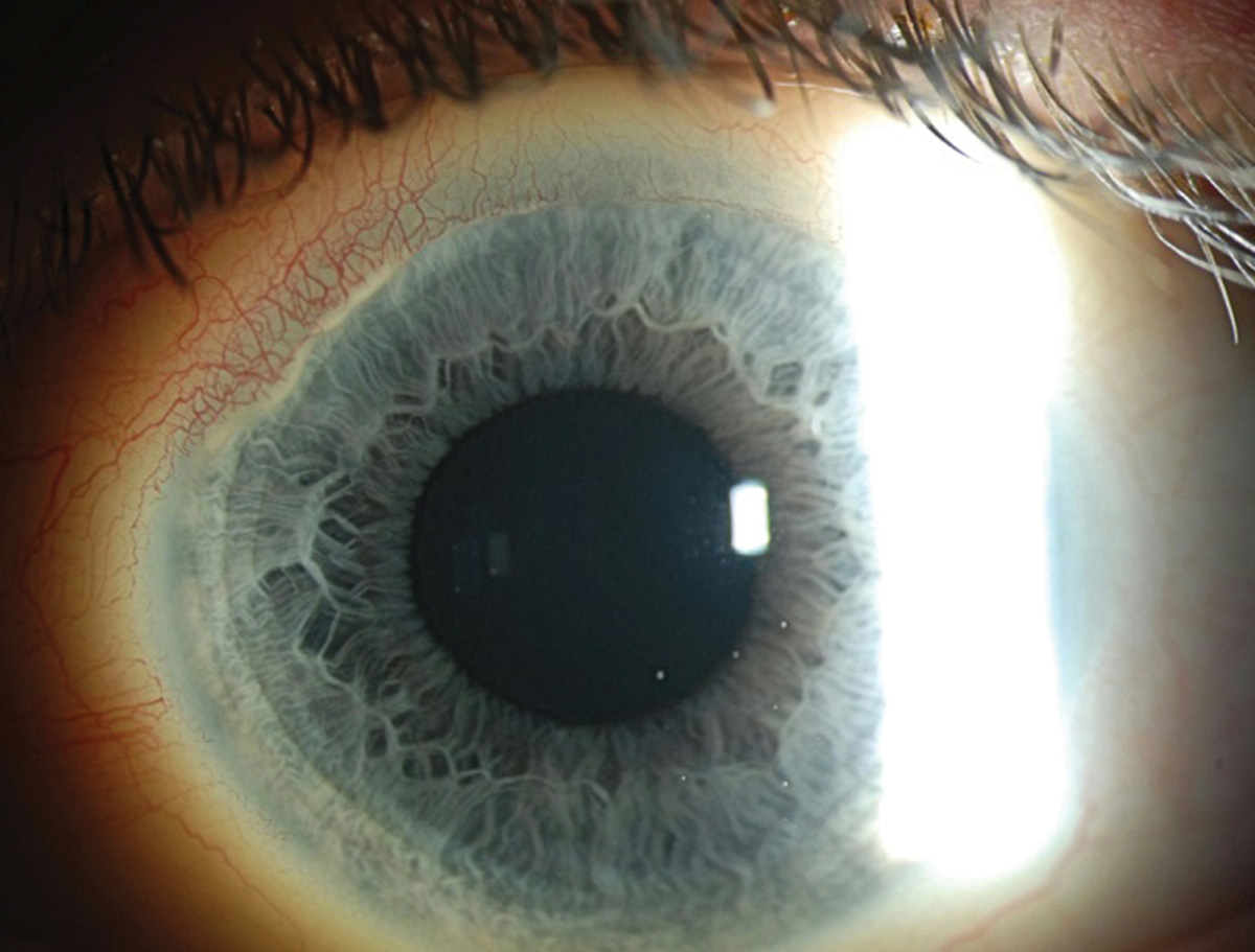
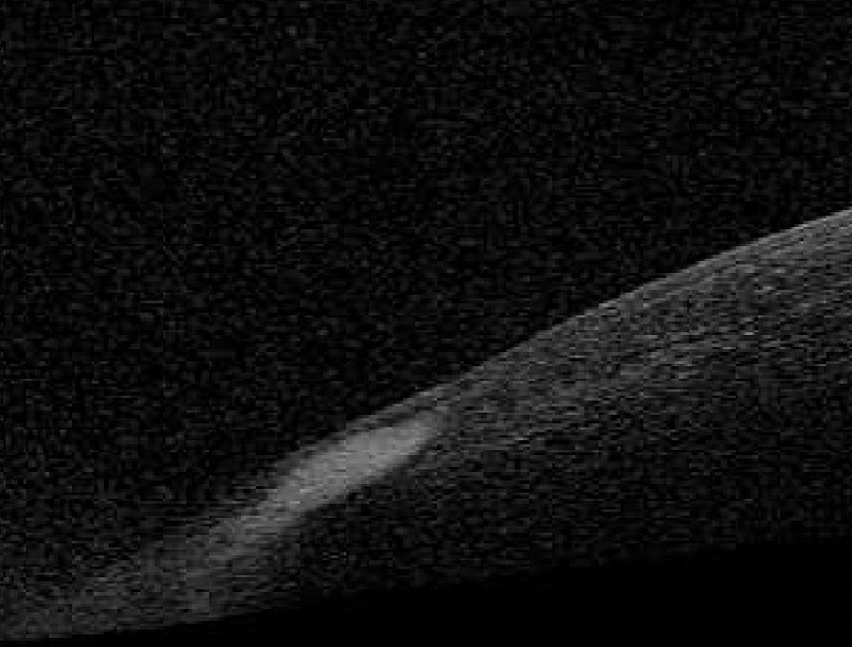
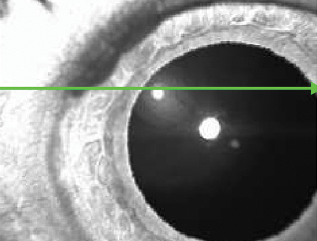
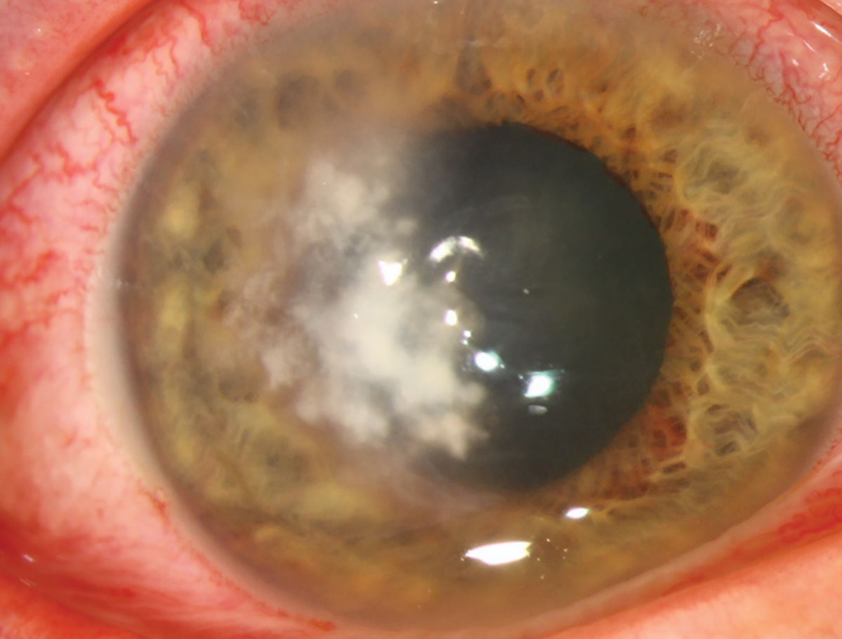
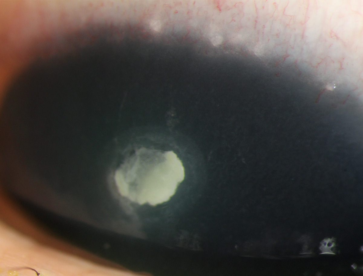
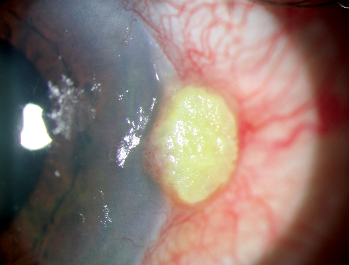
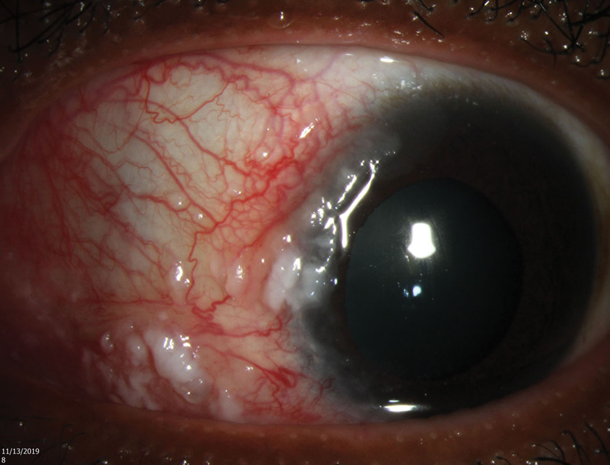
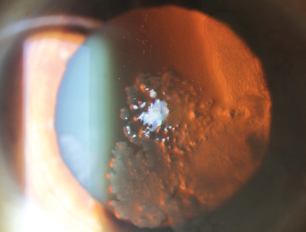
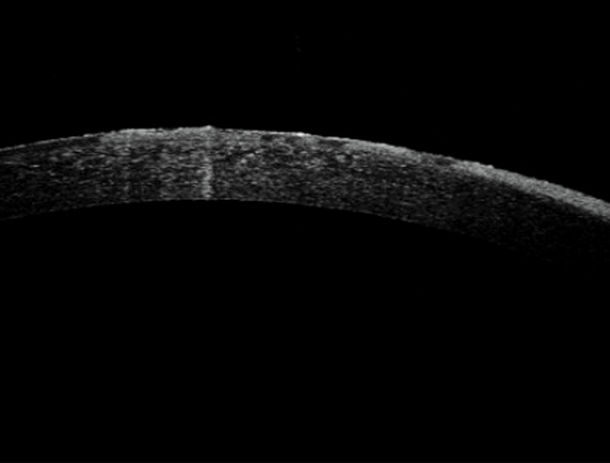
Ocular surface squamous neoplasia (OSSN) is an umbrella term for an array of abnormal growths, and lacks well-defined characteristics as a result. The left photo shows a case of leukoplakic OSSN at the limbus. The center and right images show opalescent, flat corneal lesions. The AS-OCT highlights abrupt transitions between normal and irregular epithelium despite lacking significant thickness.
Suggested reading:
A Guide to Conjunctival Tumors
Is That Conjunctival Lesion Cancerous?
When a Red Eye Prompts a Red Alert
Firework injury
 |
| Photo: University of Iowa. Click image to enlarge. |
Fireworks may cause globe rupture, chemical or thermal burns as well as corneal abrasion and retinal detachments.
Suggested reading:
Symblepharon
 |
| Photos: Alison Bozung, OD. Click image to enlarge. |
 |
| Click image to enlarge. |
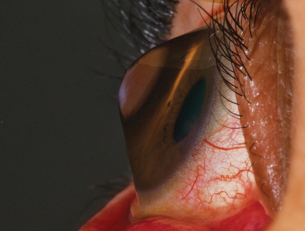
The first photo shows adherence of the bulbar and palpebral conjunctivae, or symblepharon, in a patient who would later be diagnosed with ocular cicatricial pemphigoid. The second shows severe symblepharon in an advanced case of ectodermal dysplasia.
Suggested reading:
Systemic Disease Rising to the Surface
Amelanotic melanoma
 |
| Photo: University of Iowa. Click image to enlarge. |
This is a rare subtype of melanoma where the melanocytes fail to produce melanin.
Suggested reading:
A Guide to Conjunctival Tumors
Is That Conjunctival Lesion Cancerous?
Conjunctival melanoma
 |
| Photos: University of Iowa; Carol Shields, MD. Click image to enlarge. |
 |
| Click image to enlarge. |
 |
| Click image to enlarge. |
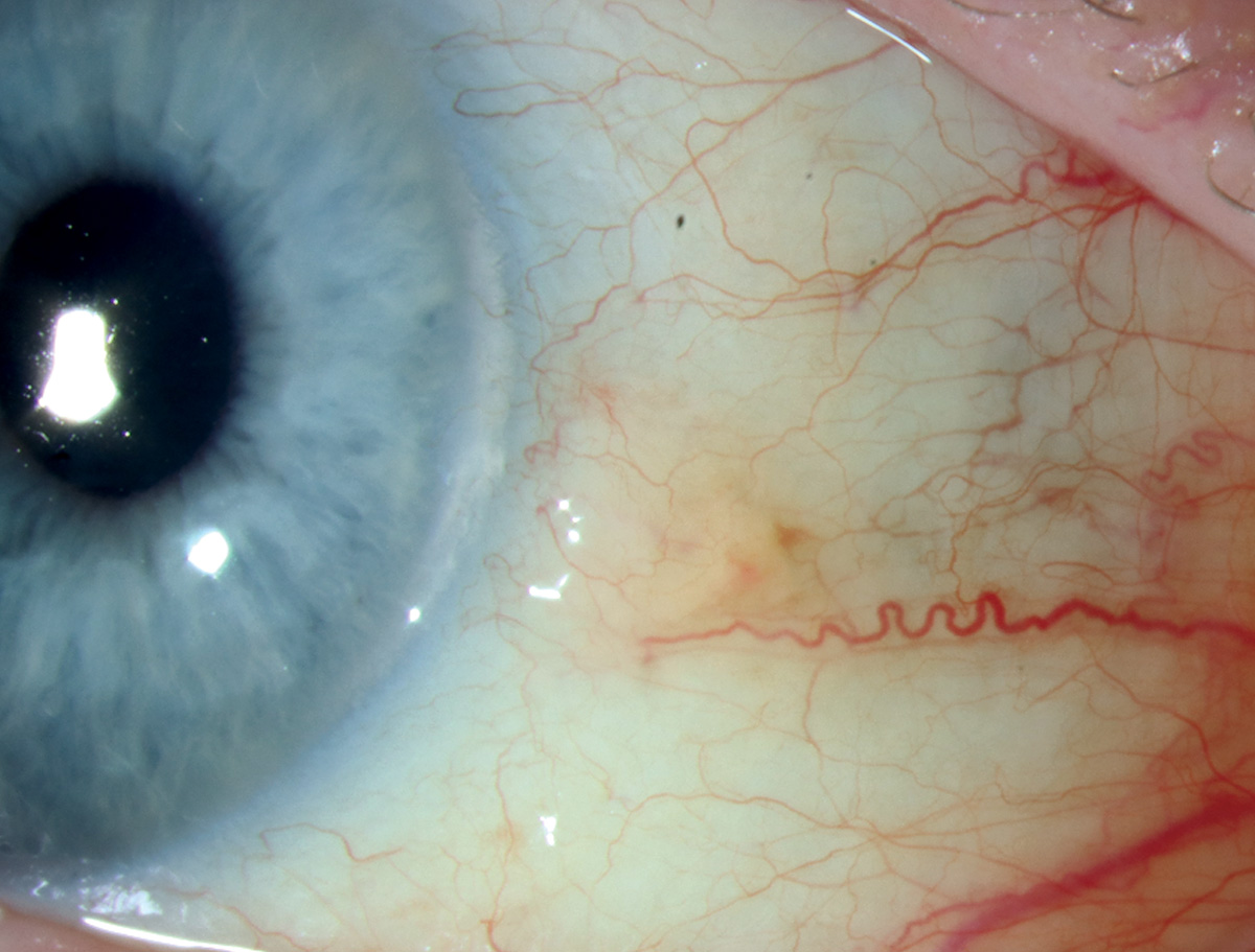
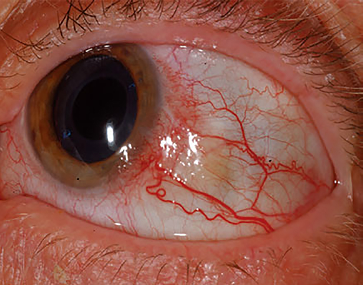
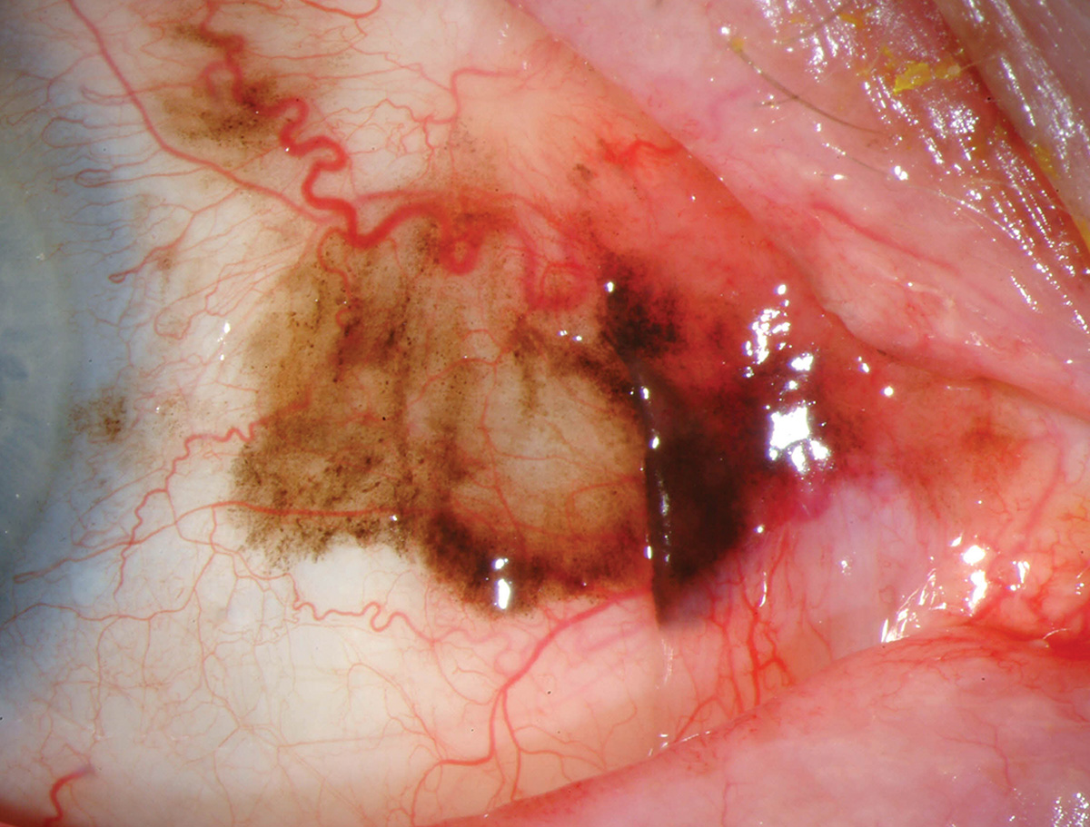
Seen in the first photo is a typical conjunctival melanoma. Often, large feeder vessels will surround the pigmented area. These lesions can also be non-pigmented (second photo) or mixed pigmented/non-pigmented (third photo).
Suggested reading:
A Guide to Conjunctival Tumors
Is That Conjunctival Lesion Cancerous?
Conjunctival chemosis
 |
| Photo: University of Iowa. Click image to enlarge. |
Severe conjunctival chemosis experienced by a burn victim who received copious fluid administration.
Suggested reading:
Distinguish The Rare Conjunctival Tumor
Stevens-Johnson syndrome
 |
| Photos: Alison Bozung, OD. Click image to enlarge. |
 |
| Click image to enlarge. |
 |
| Click image to enlarge. |
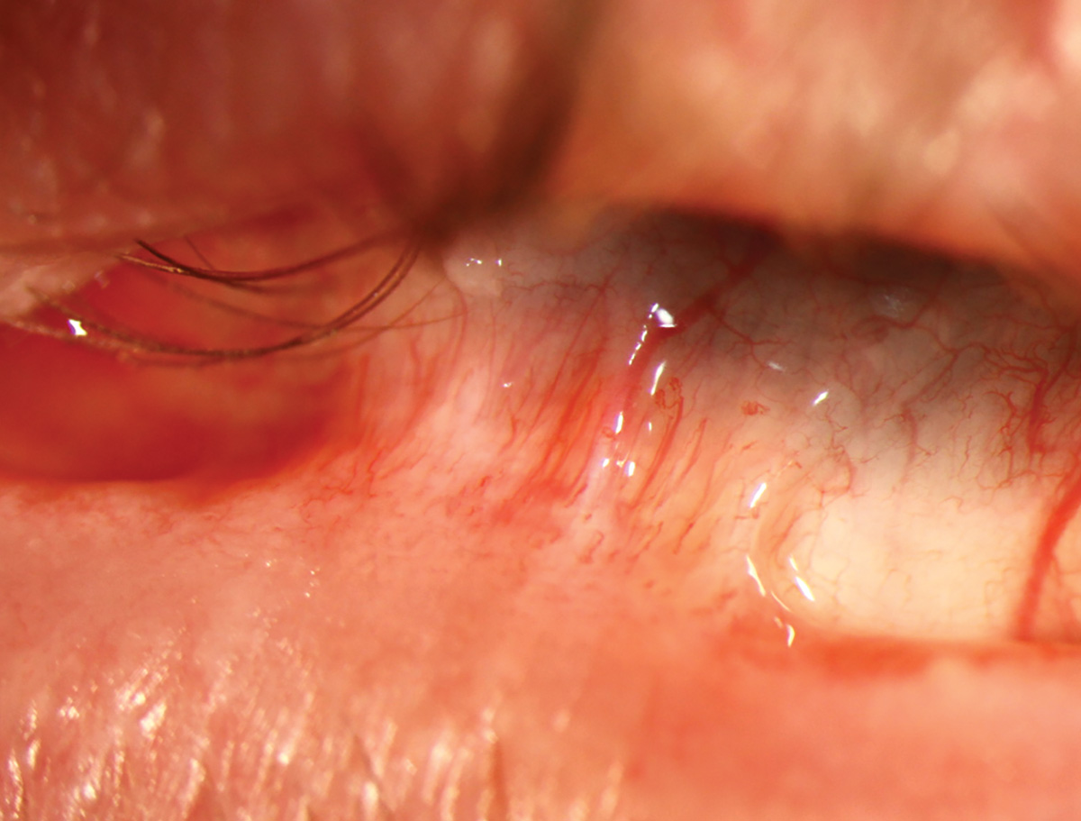
This young male presented with red, painful eyes in the setting of a recent Stevens-Johnson syndrome reaction to a medication. There is inflammatory epithelial sloughing along the mucous membranes including the mucocutaneous eyelid margins and bulbar conjunctiva. This condition may cause blindness due to ocular surface scarring and must be treated aggressively. Cases with more extensive systemic/skin involvement would be considered toxic epidermal necrolysis, a condition that can be fatal.
Epidemic keratoconjunctivitis
 |
| Photo: Alison Bozung, OD. Click image to enlarge. |
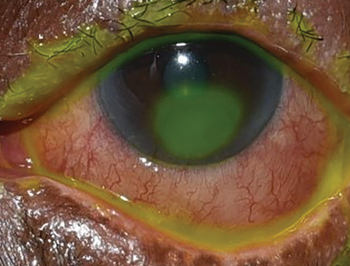
EKC is often seen with pseudomembranes (stained with NaFl here) and subepithelial infiltrates.
Suggested reading:
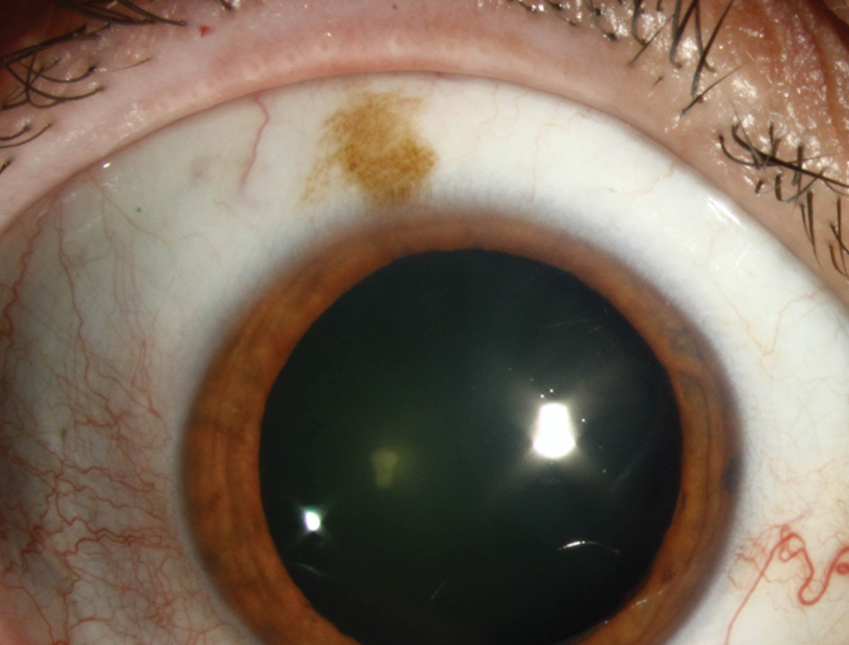
Nevus of Ota
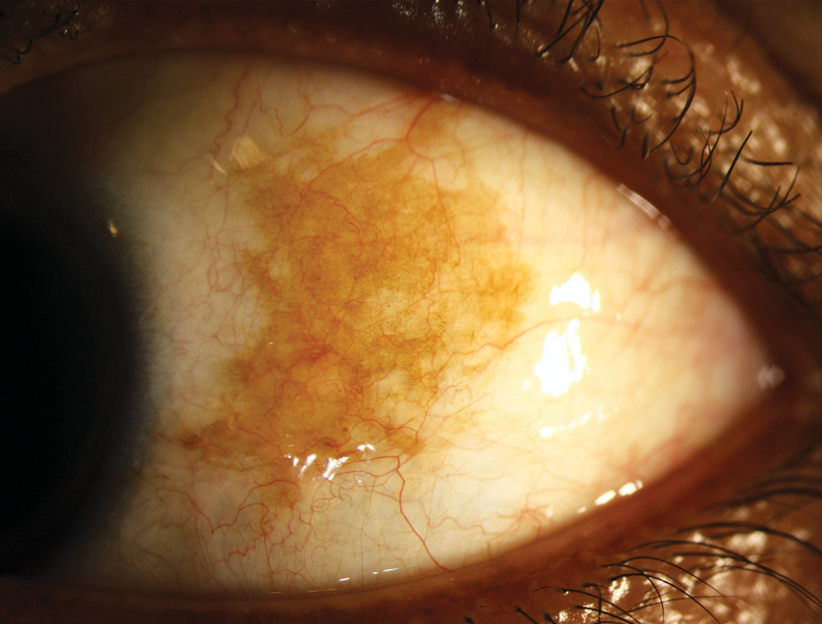 |
| Photos: University of Iowa. Click image to enlarge. |
 |
| Click image to enlarge. |
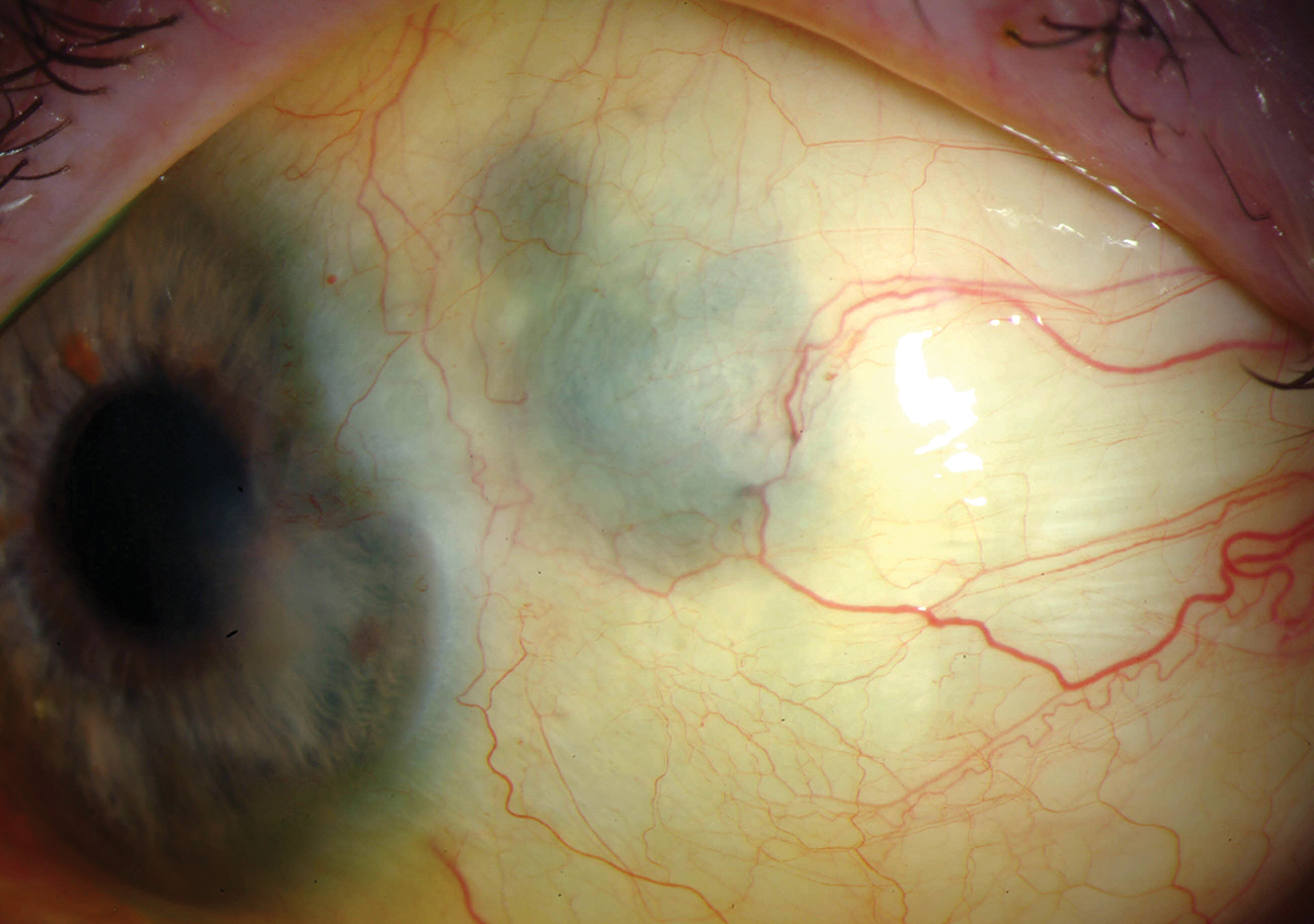
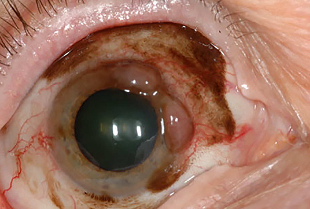
Nevus of Ota (also called oculodermal melanosis) is a congenital hyperpigmentation of the sclera. The condition is usually benign but it may have a malignant transformation. Increased suspicion for eventual glaucoma development in these cases is also appropriate.
Suggested reading:
A Guide to Conjunctival Tumors
Iris sphincter tear
 |
| Photo: Alison Bozung, OD. Click image to enlarge. |
Iris sphincter tears from blunt trauma may cause an irregular pupillary margin and permanent dilation.
Suggested reading:
Pupil Testing: Implications for Diagnosis
Iridodialysis
 |
| Photo: Alison Bozung, OD. Click image to enlarge. |
In iridodialysis, the iris separates from the scleral spur. This patient was involved in a motor vehicle accident.
Suggested reading:
Traumatic pupil
 |
| Photo: University of Iowa. Click image to enlarge. |
Traumatic pupil represents a traumatic mydriasis that occurs in response to blunt force trauma.
Suggested reading:
Angle recession
 |
| Photos: University of Iowa. Click image to enlarge. |
 |
| Click image to enlarge. |
Development of angle recession following blunt ocular trauma may cause glaucoma days or years after the injury.
Suggested reading:
Gonioscopy: A Simple Tool, Too Often Forgotten
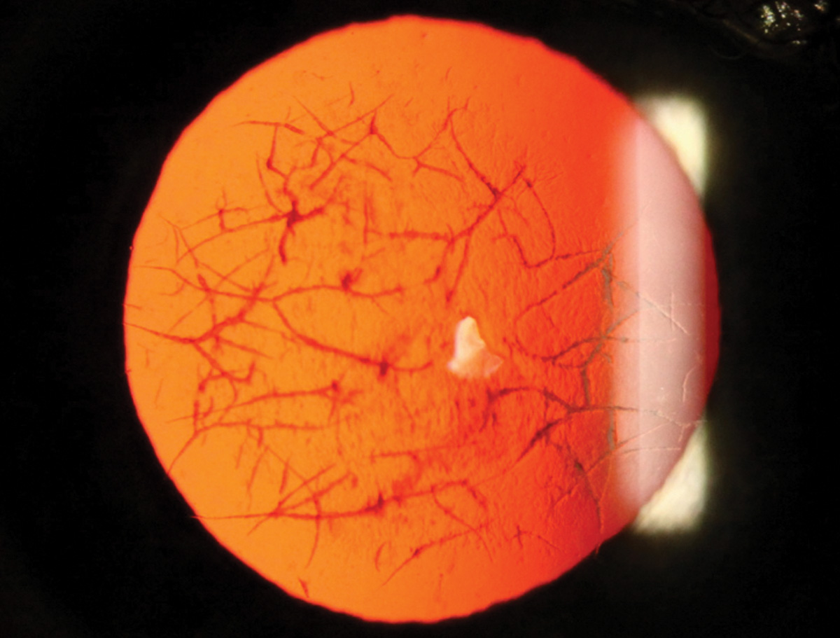
Rubeosis iridis
 |
| Photos: Mohammad Rafieetary, OD. Click image to enlarge. |
 |
| Click image to enlarge. |
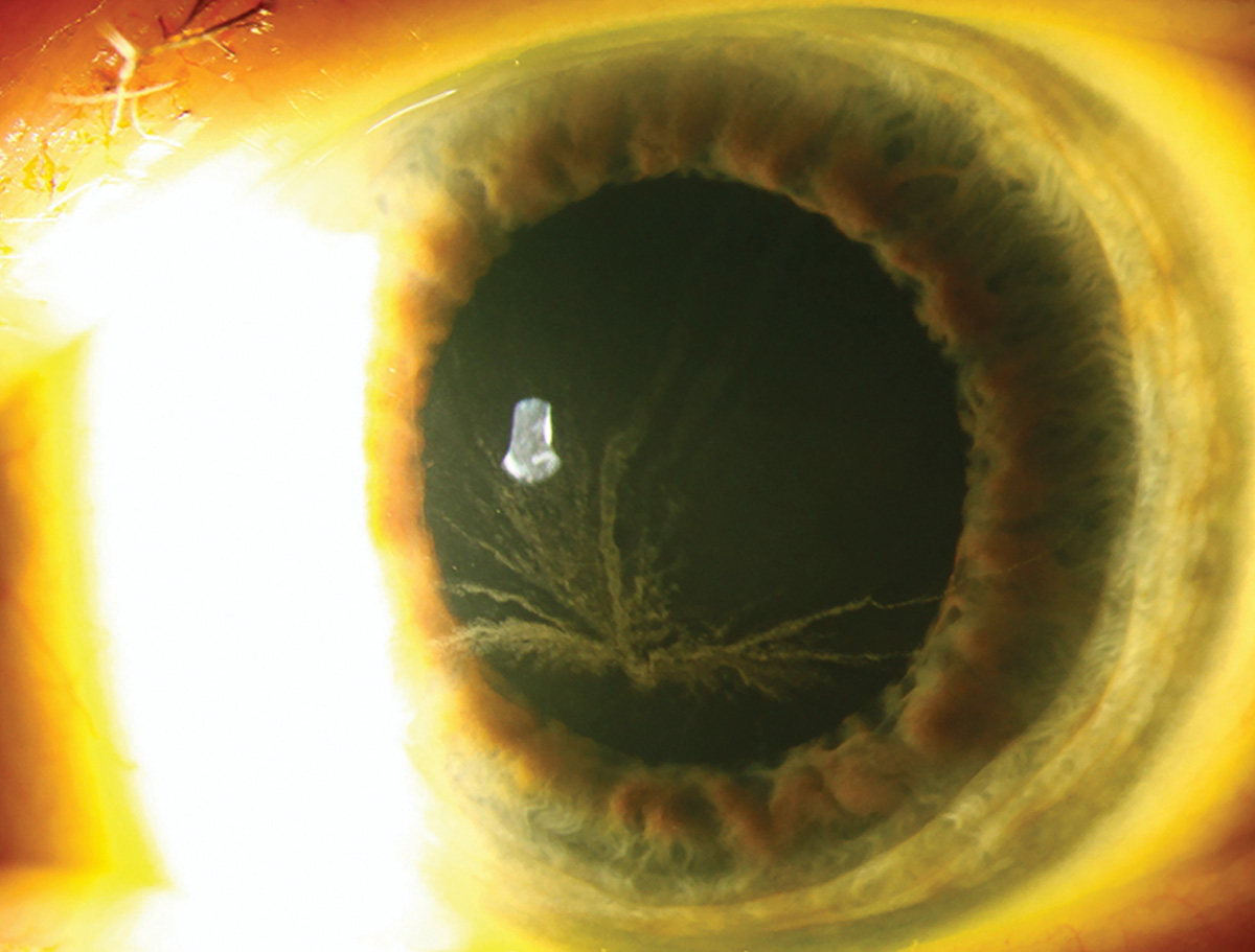
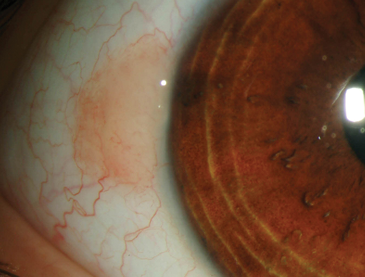
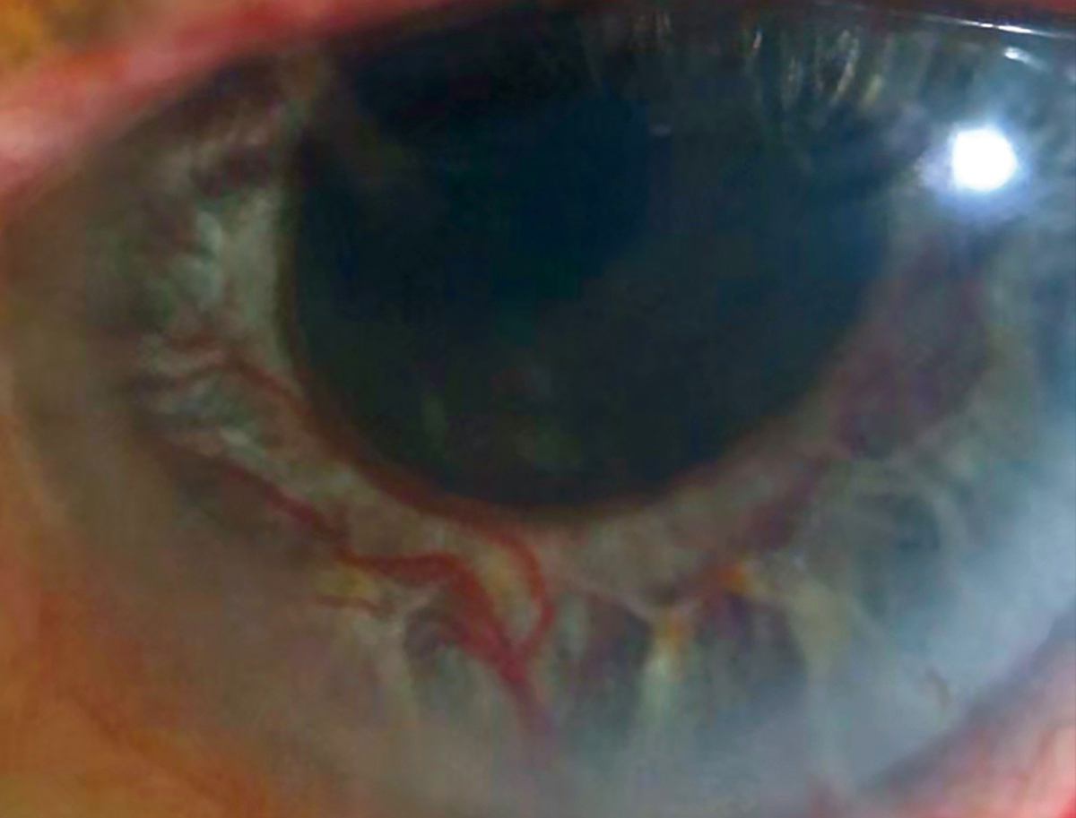
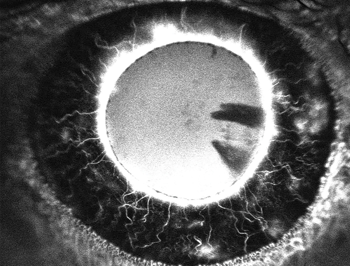
Rubeosis iridis requires careful evaluation to detect, as it may present very subtly, often initially at the pupillary margin. Gonioscopy may detect neovascularization in the angle. IVFA of the iris may be helpful to show hyperfluorescence from the irregular vasculature.
Suggested reading:
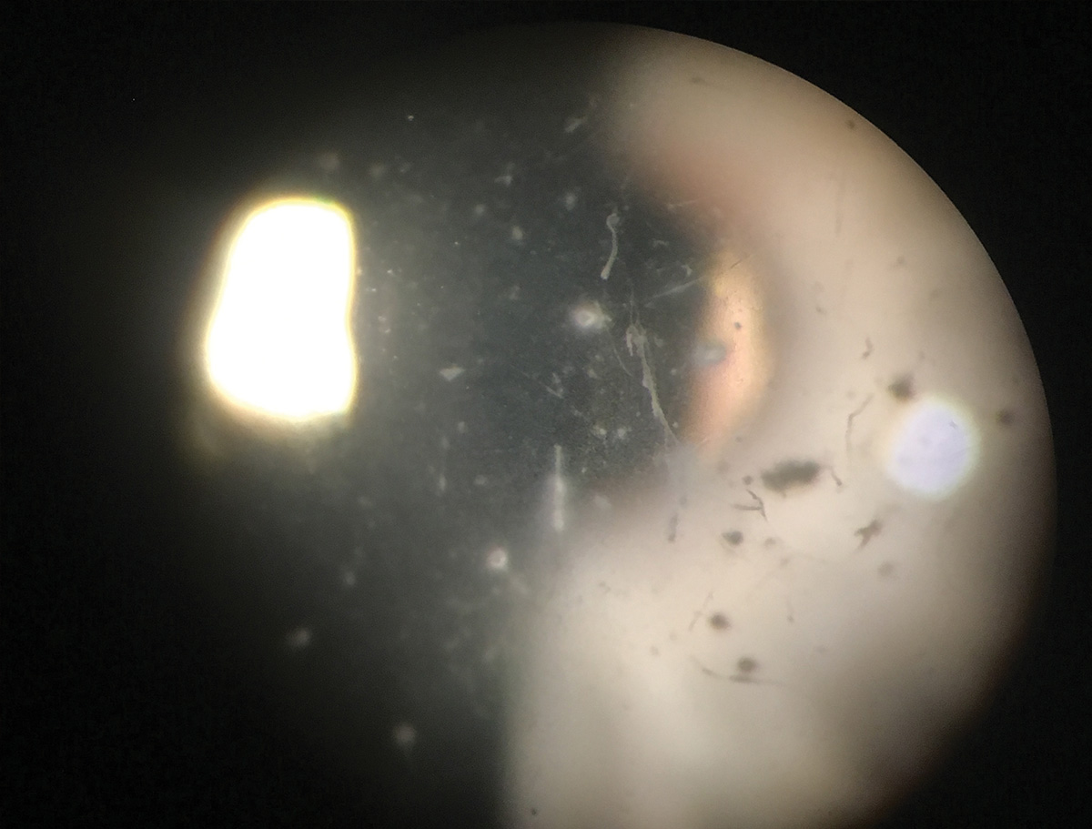
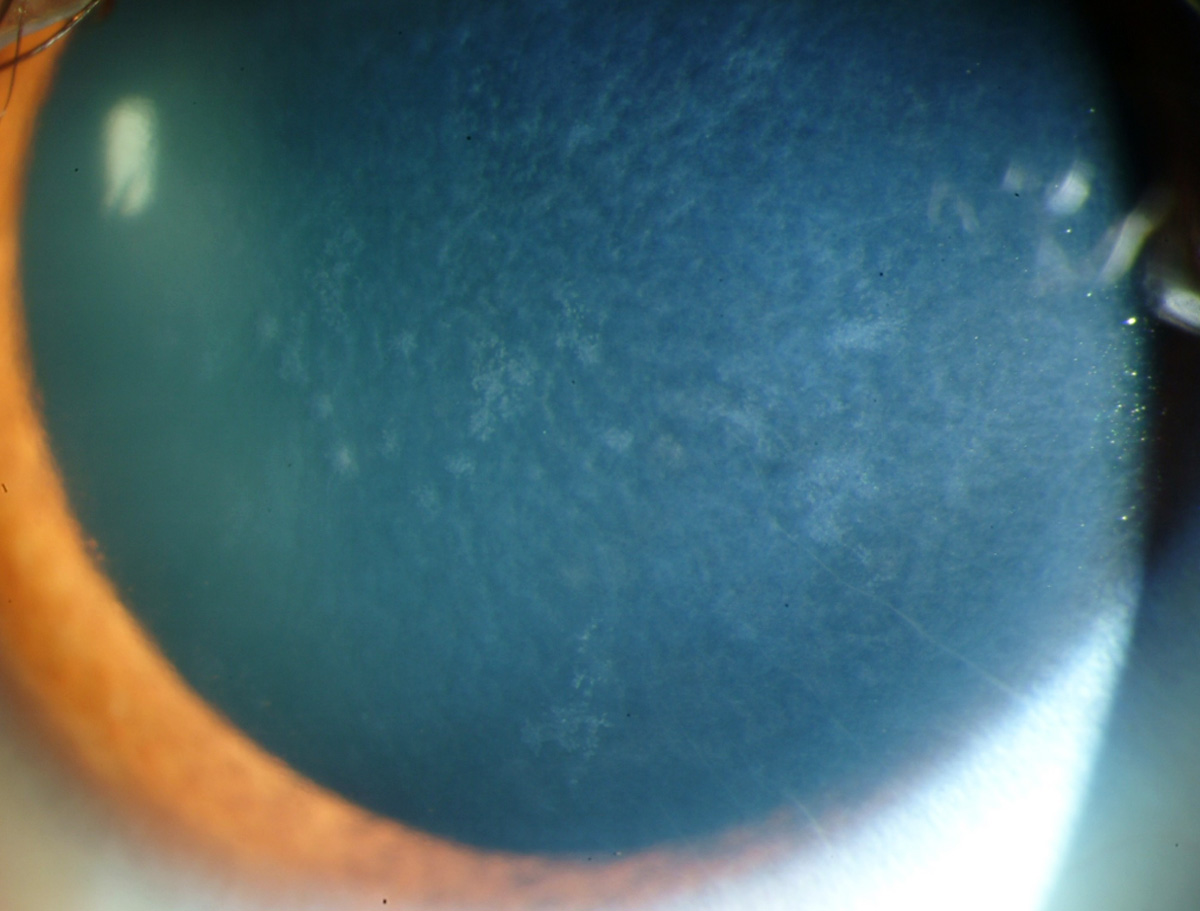
Keratic precipitates
 |
| Photo: University of Iowa. Click image to enlarge. |
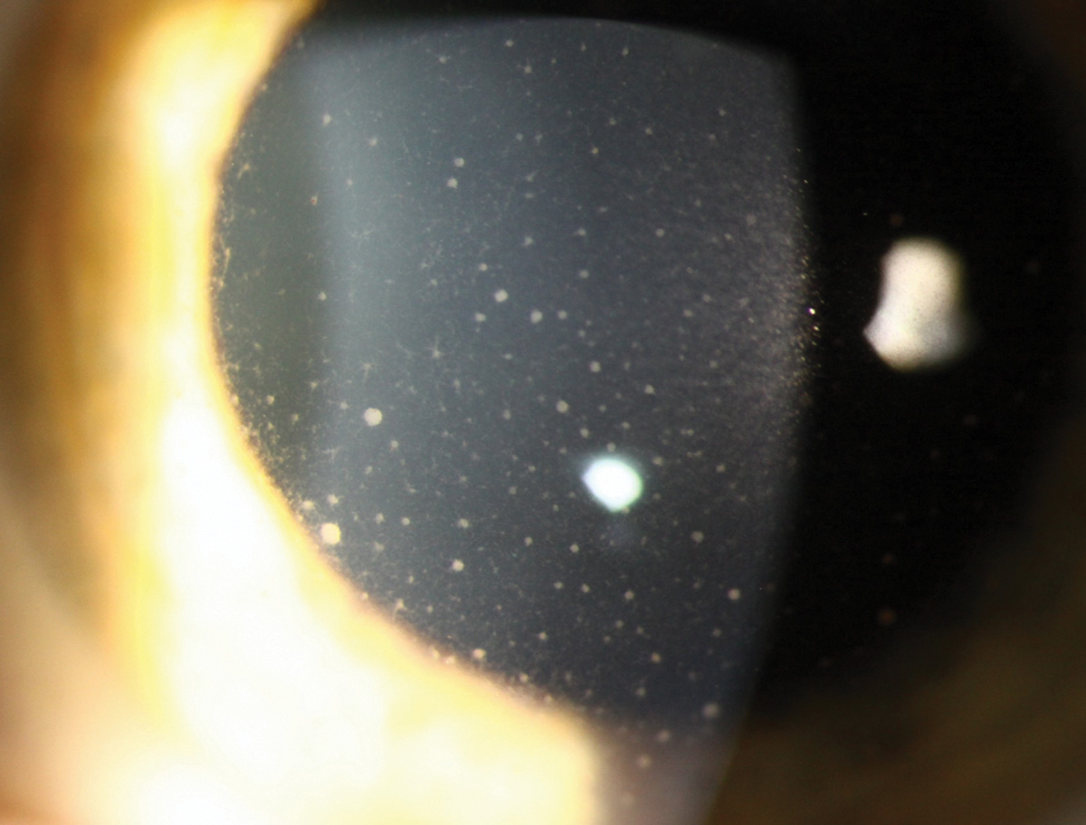
Keratic precipitates in iridocyclitis are polymorphonuclear cells and lymphocytes located on the corneal endothelium.
Suggested reading:
Practical Pearls for Managing Anterior Uveitis
Glaucomatocyclitic Crisis: A Not-So-Benign Disease?
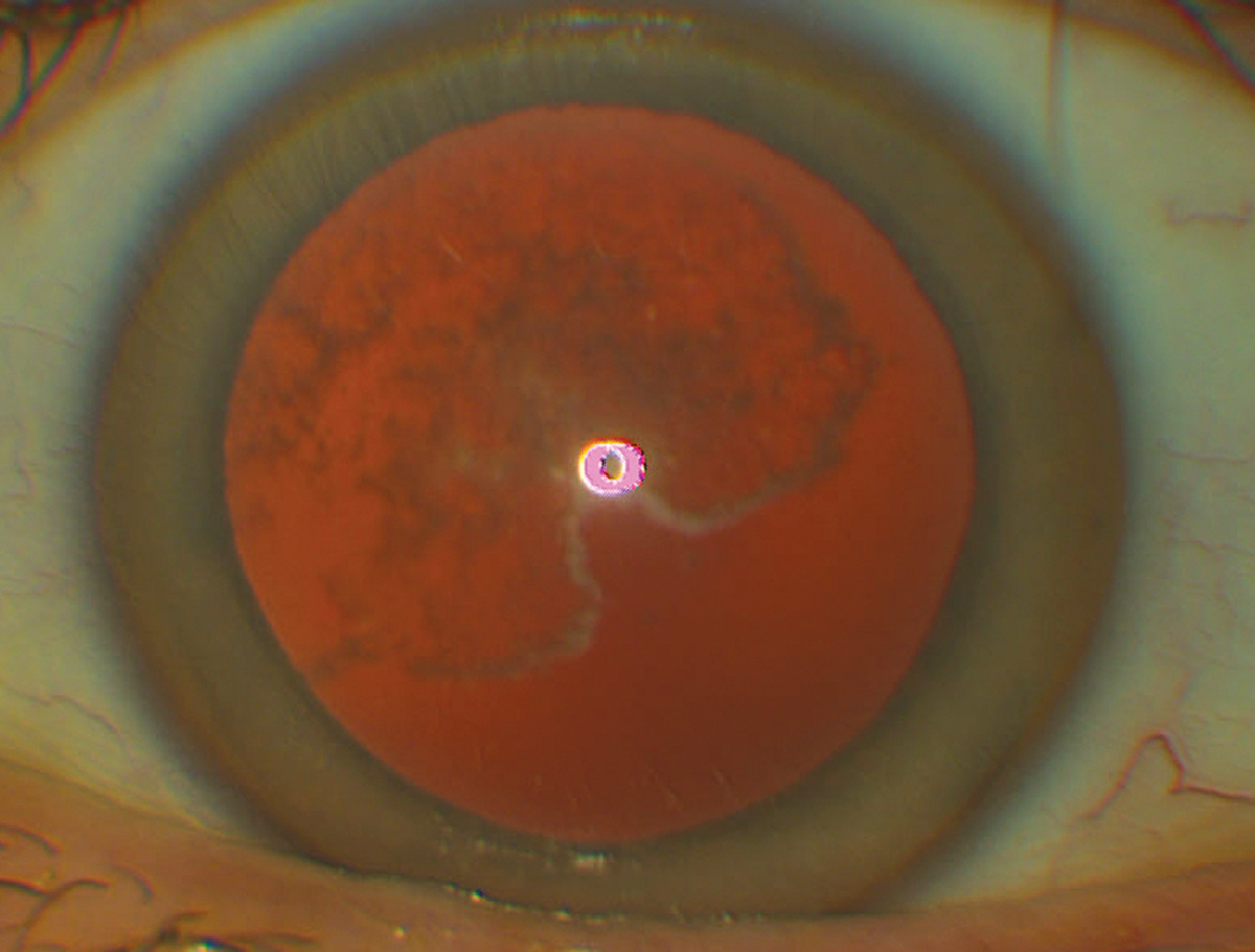
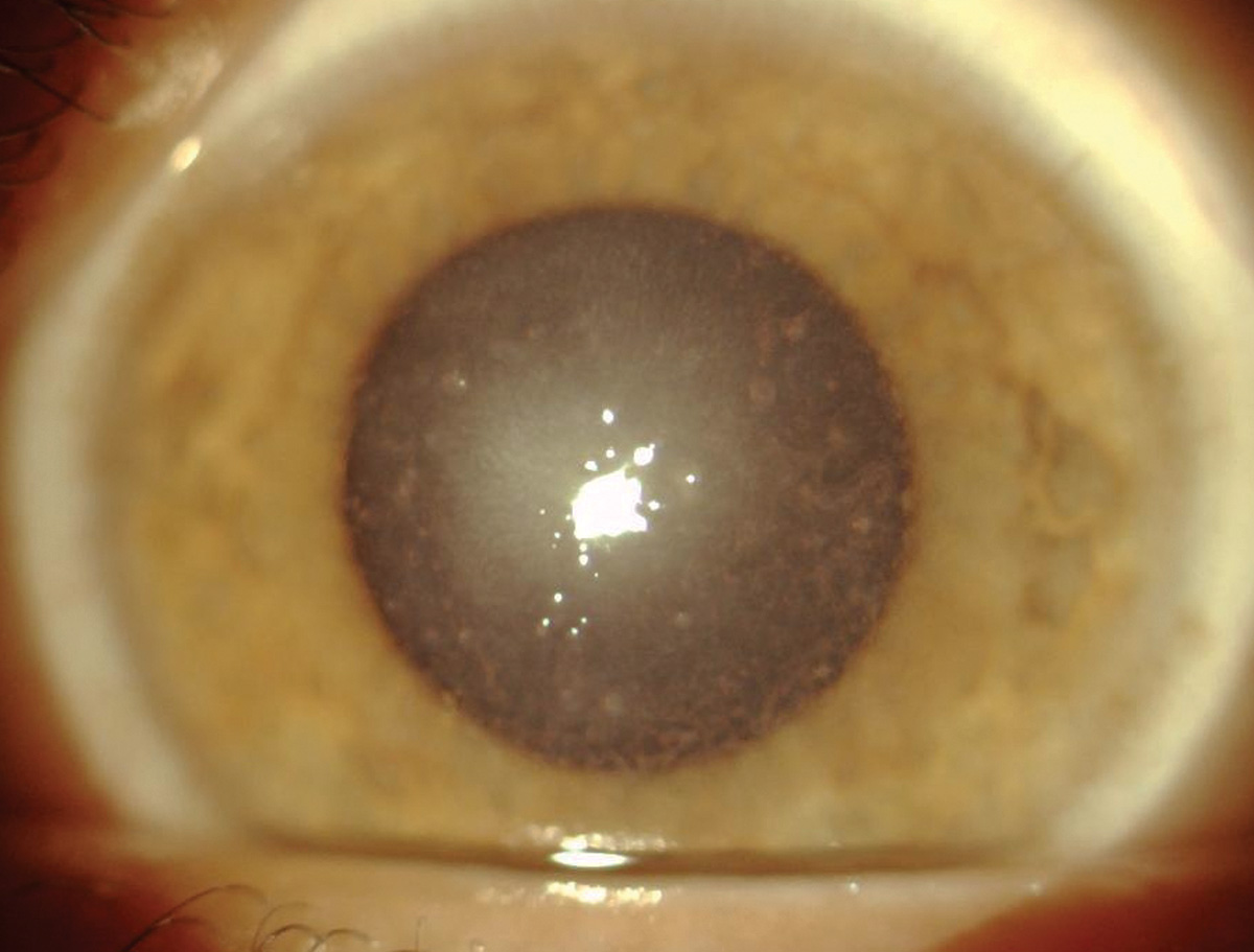
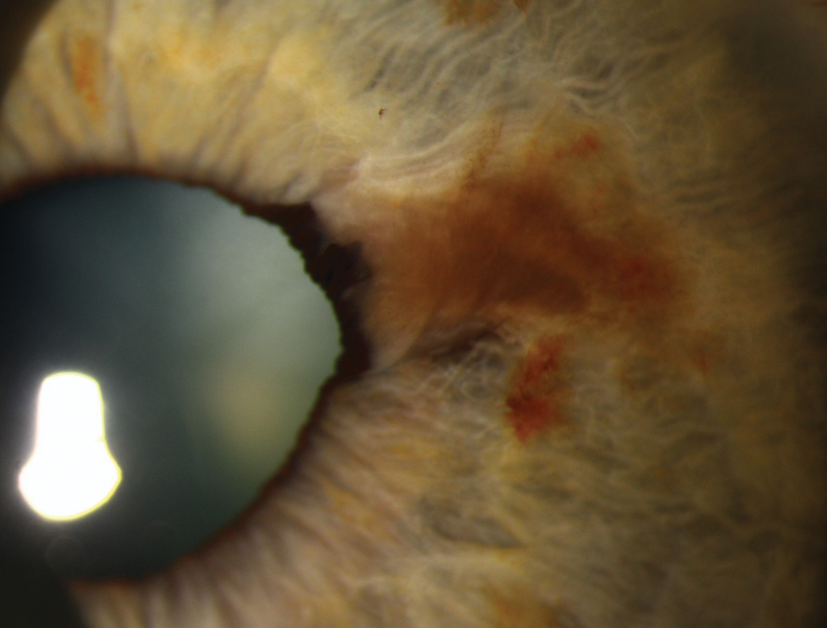
Iris melanocytosis
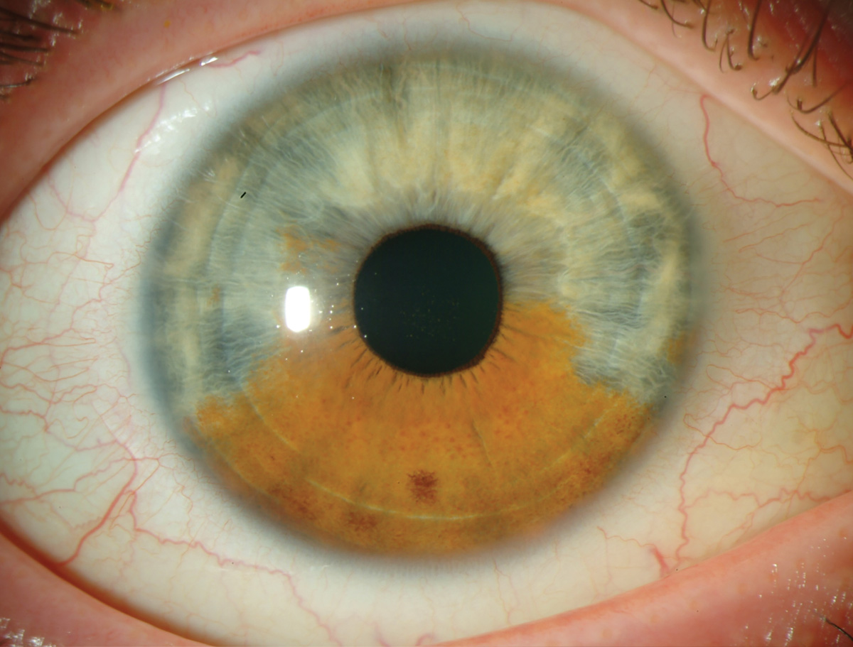 |
| Photo: University of Iowa. Click image to enlarge. |
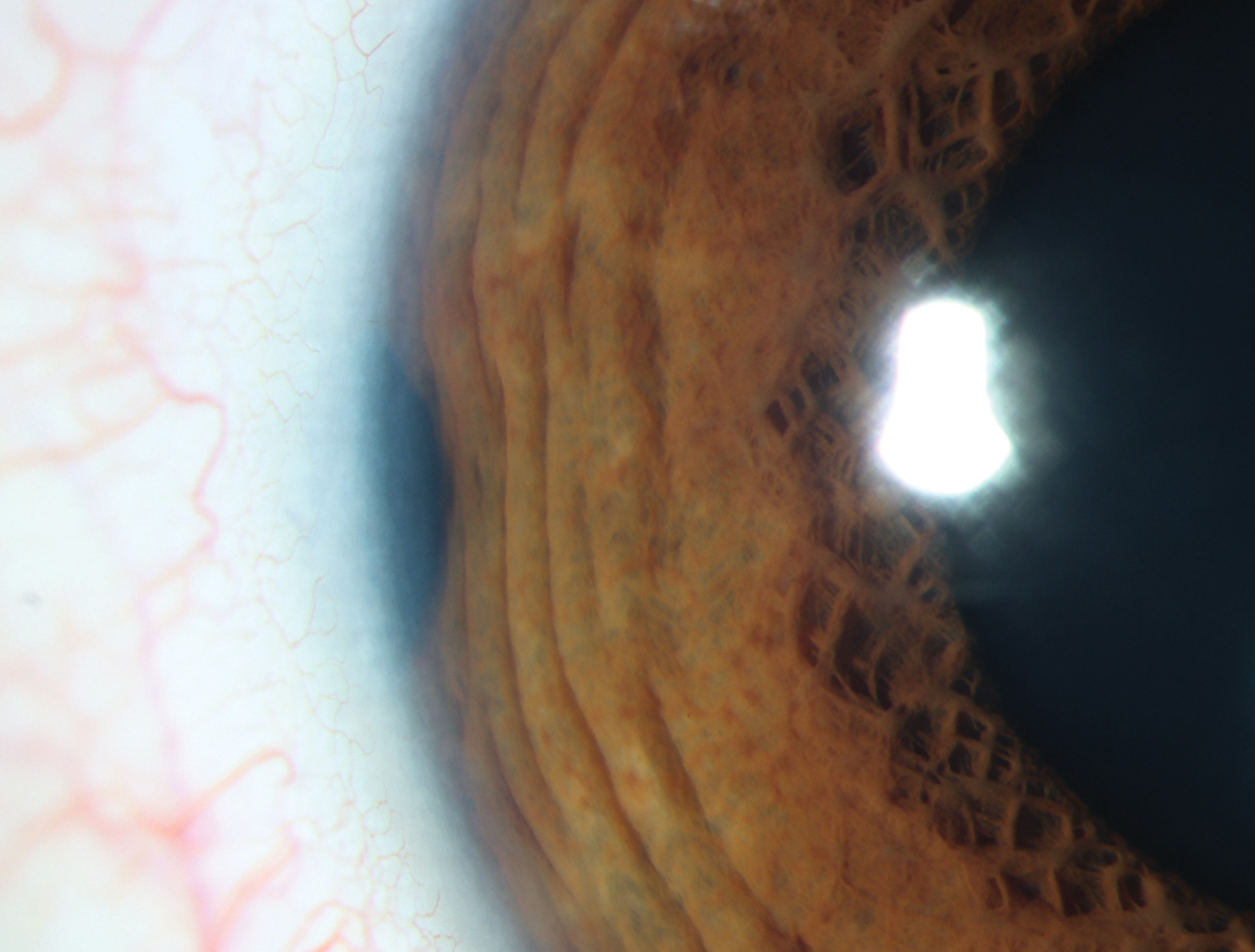
Though usually considered benign, iris melanocytosis carries an increased lifetime risk for melanoma.
Suggested reading:
Earlier Detection of Iris Melanocytic Tumors Needed
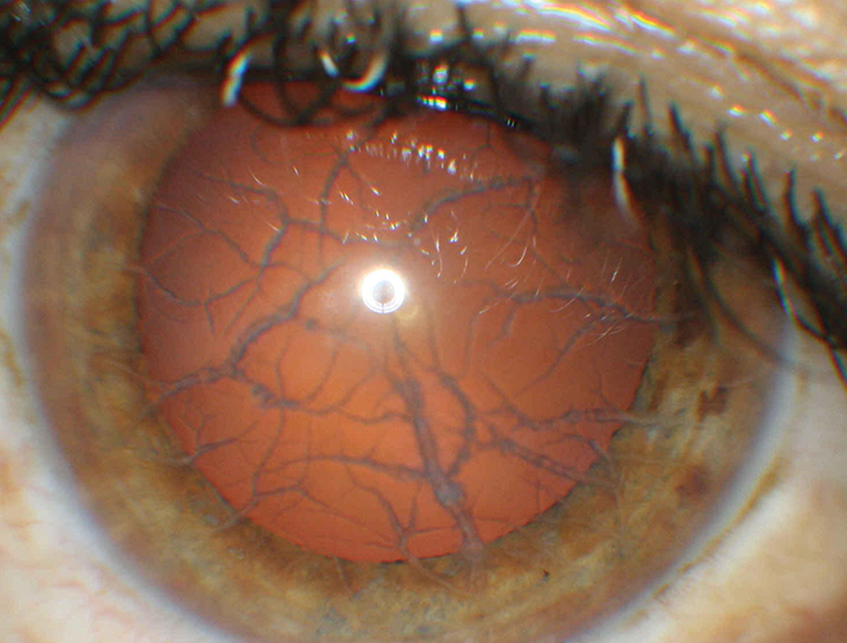
Ectropion uvea
 |
|
Photo: University of Iowa. Click image to enlarge. |
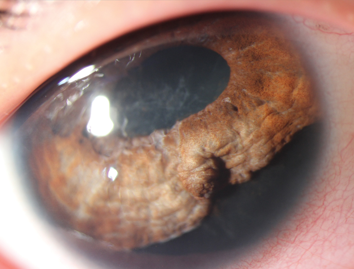
Ectropion uvea may be acquired or congenital. Acquired cases may rise from inflammation, ischemia or neoplasm.
Suggested reading:
